小腿肌肉mri解剖结构图

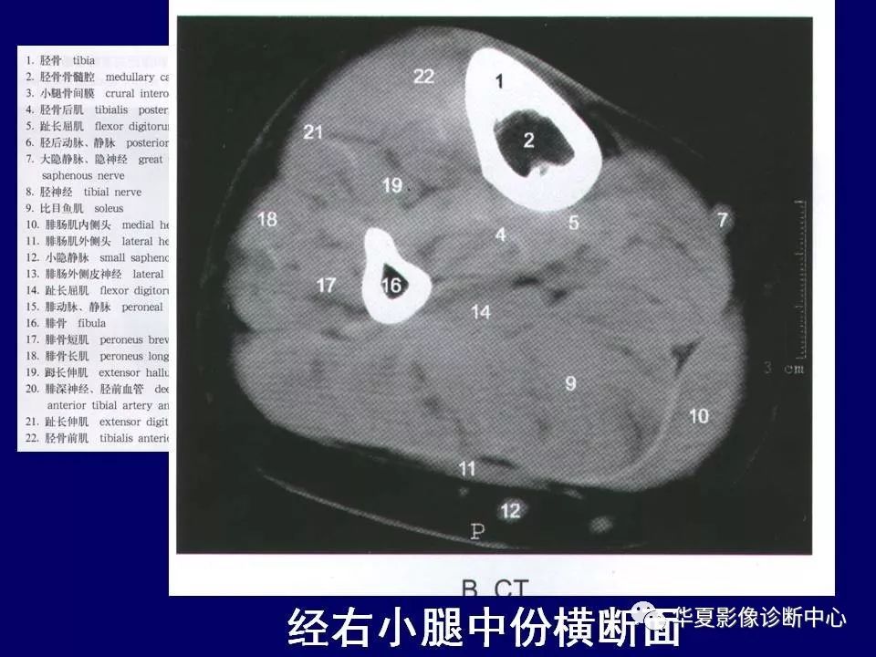
下肢的断层解剖
图片尺寸960x720
小腿部断层解剖 详细标注_全网_影像_系统
图片尺寸640x534
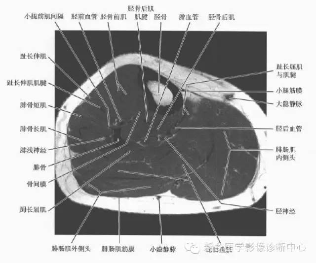
小腿部断层解剖详细标注
图片尺寸640x539
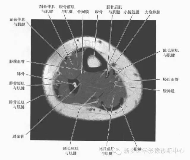
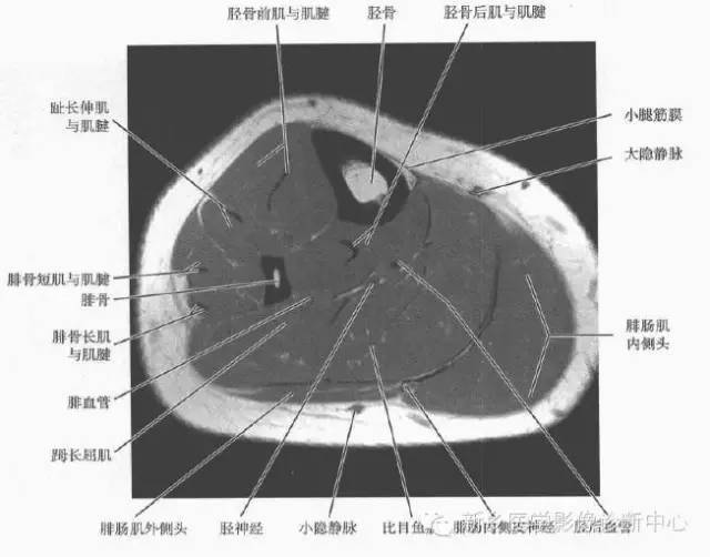
小腿部断层解剖详细标注
图片尺寸640x502关节影像膝关节的磁共振mri解剖表现
图片尺寸853x564
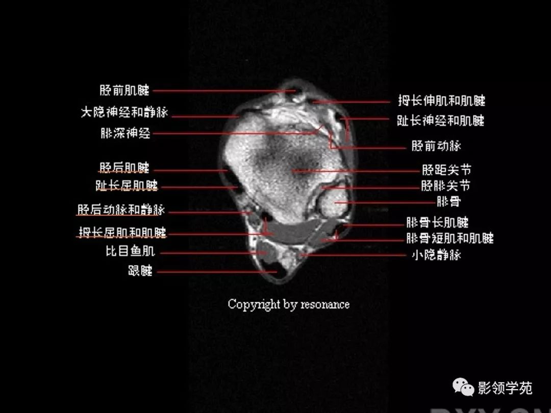
踝关节正常mri解剖图谱
图片尺寸570x500如何准确找到韧带:骨性解剖结构标志7272 冠状面韧带观察7272
图片尺寸690x1036
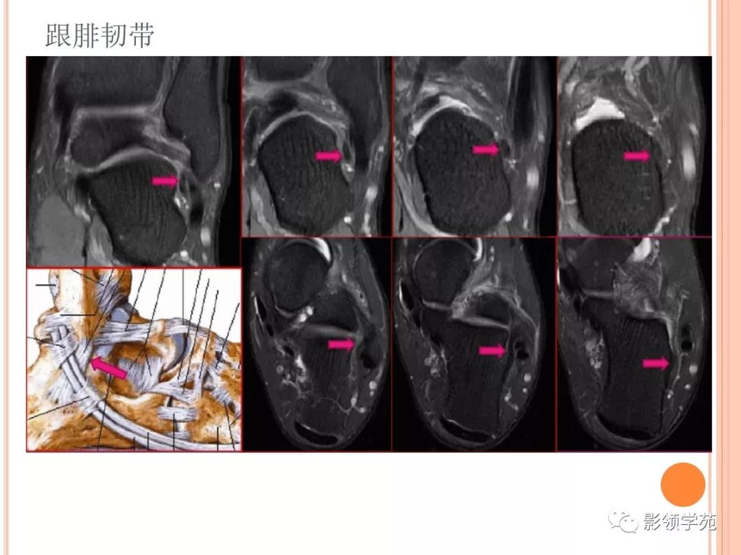
踝关节解剖及损伤mri诊断
图片尺寸1080x608最全的下肢mri解剖
图片尺寸960x720
左小腿磁共振显示骨折端无明显炎症信号.
图片尺寸2000x2637
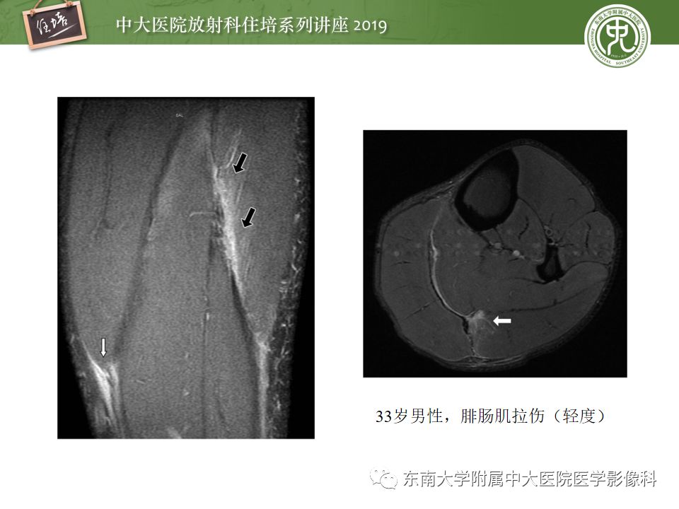
【中大放射住培系列讲座】创伤性肌肉病变的mr诊断
图片尺寸960x720
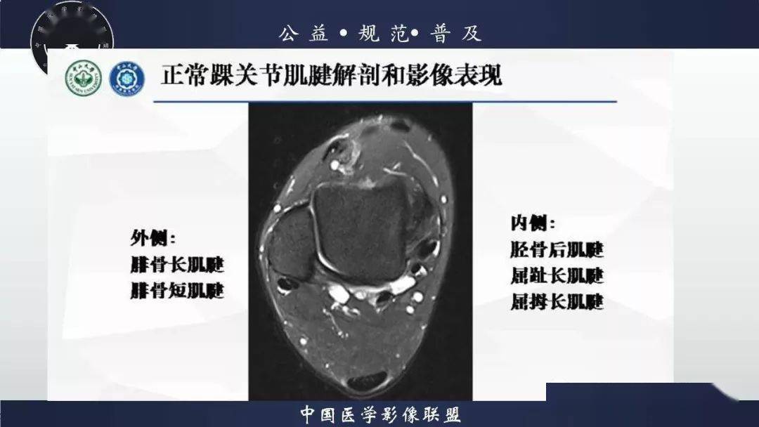
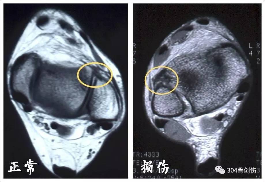
踝关节mri解剖和常见损伤类型
图片尺寸1080x810
踝关节mri解剖和常见损伤类型
图片尺寸1080x810
②前后位片上胫腓骨重叠>6mm或大于腓骨宽度的42%;mri能够直观看到下
图片尺寸919x634
值得收藏 | 踝关节mri的断层解剖_网易订阅
图片尺寸660x732
正常膝关节mri解剖图谱
图片尺寸570x336下面这张是小腿上部横截面图
图片尺寸357x212
膝关节mri解剖ppt
图片尺寸1080x810最全的下肢mri解剖
图片尺寸960x720
精美膝关节mr解剖图谱-建议收藏!
图片尺寸743x767


猜你喜欢:小腿肌肉mri解剖图小腿肌肉mr横断解剖图小腿mri断层解剖图小腿解剖结构图大腿肌肉解剖mri小腿肌肉断层解剖图大腿肌肉mr横断解剖图腓肠肌mri解剖图小腿磁共振解剖图谱下肢mri解剖图心脏的解剖结构图讲解小腿肌解剖图人体肌肉结构图示意图骨盆的解剖结构图大腿肌肉断层解剖图谱颅骨解剖结构图纵隔解剖结构图膝关节解剖结构图高清踝关节mri解剖图小腿解剖图谱大腿断层MRI解剖图垂体解剖结构mri口腔解剖图结构图面听神经mri解剖图垂体柄mri解剖图齿状核解剖mri图眼球解剖结构图呼吸系统解剖结构图正常垂体mri解剖图颈动脉解剖结构图狮子的开花像喇叭的花大全图tomford眼影03妖艳女头红色泰德发球机爆破作业人员许可证邯黄铁路2020年正式工电视机logo图片大全集裴珠泫肉白咲花图片昆明花logo郑州火车站内部分布图