左肺舌叶

两肺下叶向上延伸至t3棘突水平(图5),因此,背部除最上方区域外的所有
图片尺寸768x595
肺肿瘤影像学诊断ppt
图片尺寸1080x810
左肺上叶舌段肺癌——空洞
图片尺寸1080x810
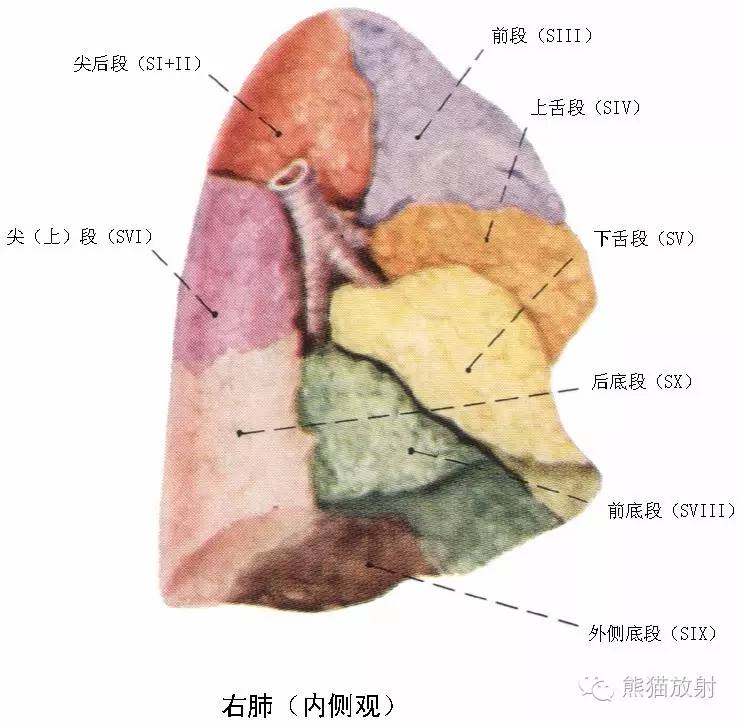
s9:外基底段,s10:后基底段】左肺上叶【s1 2:尖后段,s3:前段,s4:舌叶
图片尺寸714x711
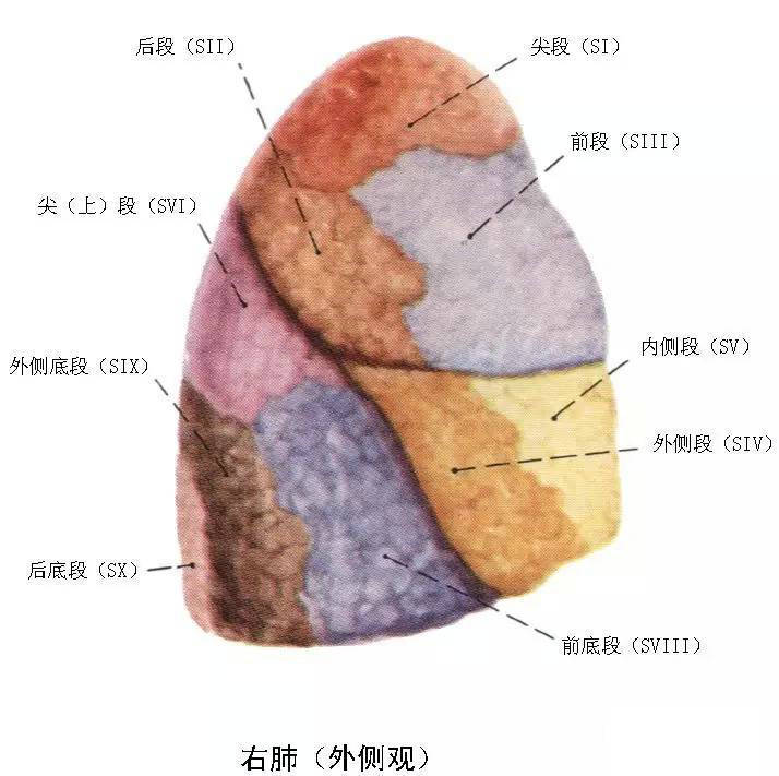
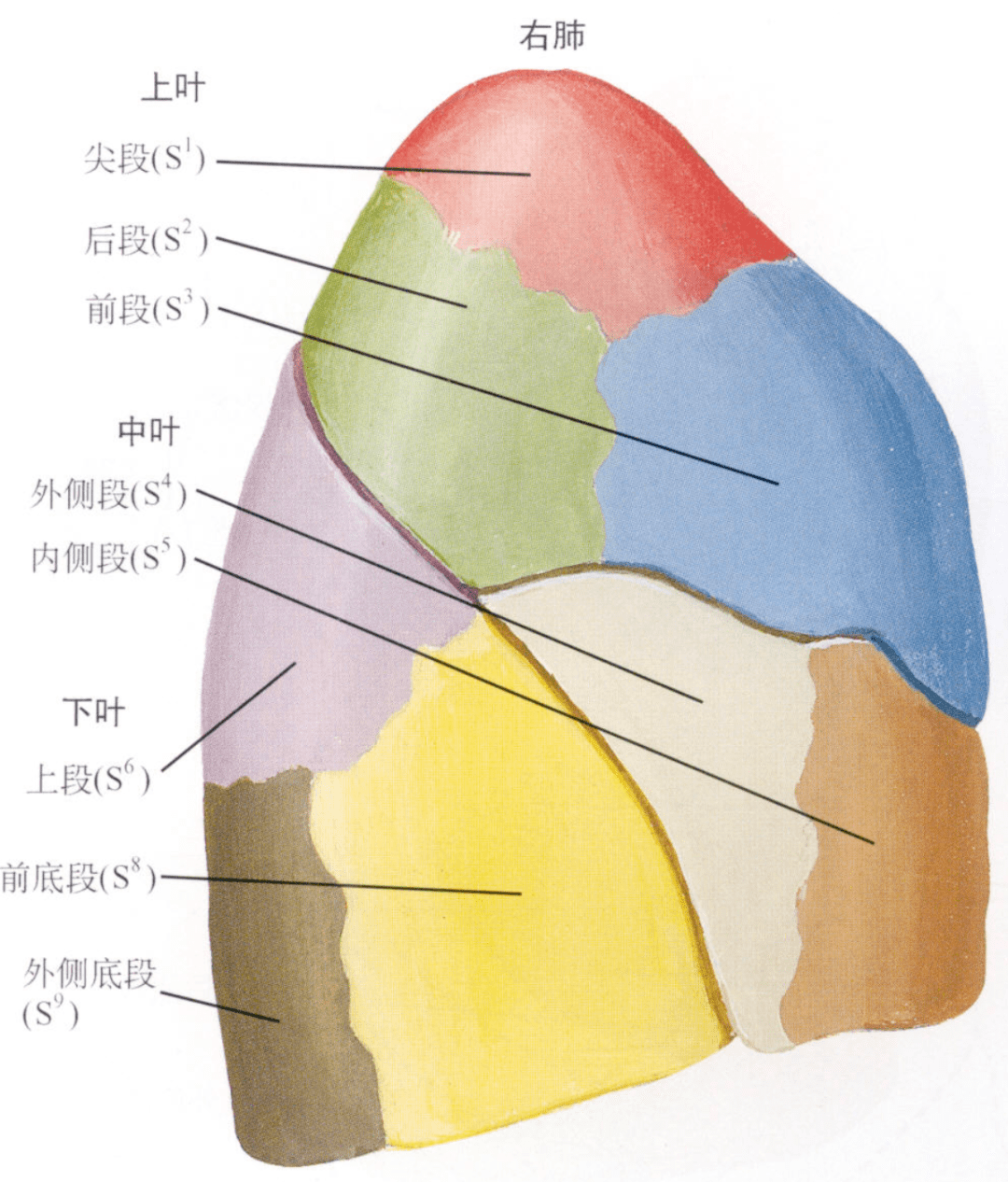
肺的分段左肺:上叶分前段,尖后段,舌段(上舌段和下舌段);下叶分上段
图片尺寸1080x810
难者不会会者不难之庖丁解肺丨一页手册协和八
图片尺寸1080x726
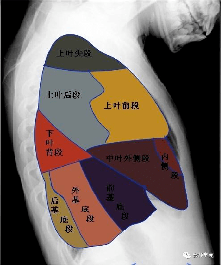
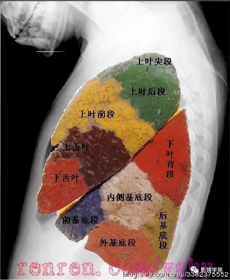
两下叶支气管分出基底段时观察中叶,舌叶及两肺下叶各基底段基底干中
图片尺寸834x829
肺脏疾病
图片尺寸500x539
呼吸系统精品解剖图收藏
图片尺寸741x724
难者不会会者不难之庖丁解肺丨一页手册协和八
图片尺寸1080x1266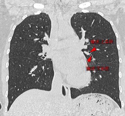
冠状面:左肺上叶上舌段和下舌段.
图片尺寸412x383
两下叶支气管分出基底段时观察中叶,舌叶及两肺下叶各基底段基底干中
图片尺寸751x901
右肺动脉 4.下后肺静脉干5.右下肺动脉 6.肺门角 7.中间支气管
图片尺寸600x422
两下叶支气管分出基底段时观察中叶,舌叶及两肺下叶各基底段基底干中
图片尺寸748x912
片示:双侧胸廓对称,肋骨走行规则,左肺上叶下舌段可见一厚壁空洞影
图片尺寸1496x1992
两肺下叶向上延伸至t3棘突水平(图5),因此,背部除最上方区域外的所有
图片尺寸768x440
人体左肺解剖示意图-人体解剖图
图片尺寸607x466
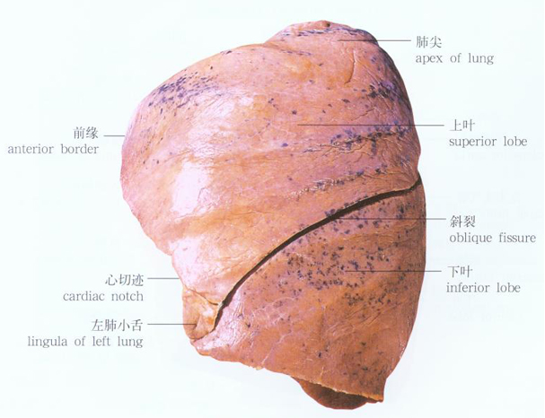
图6-11 左肺 (内面)-外科学-医学
图片尺寸950x945
内有右上肺静脉的断面,此层面能看到右肺中叶,左上肺舌叶及两肺下叶背
图片尺寸1464x854
内有右上肺静脉的断面,此层面能看到右肺中叶,左上肺舌叶及两肺下叶背
图片尺寸1384x716