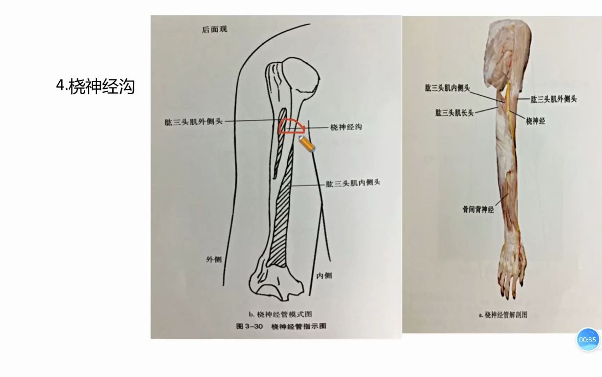
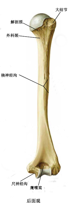
桡神经沟位于
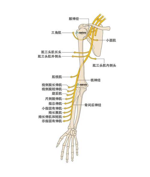
桡神经应用解剖
图片尺寸1080x1271
午休趴着睡警惕桡神经损伤
图片尺寸1080x819
桡神经易损伤点桡神经沟解析
图片尺寸1228x768
桡神经损伤
图片尺寸369x748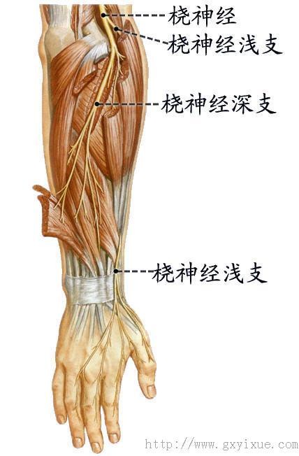
桡神经.jpg
图片尺寸428x652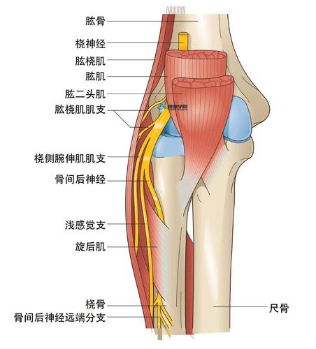
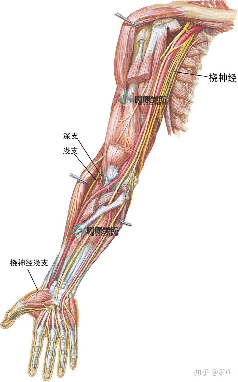
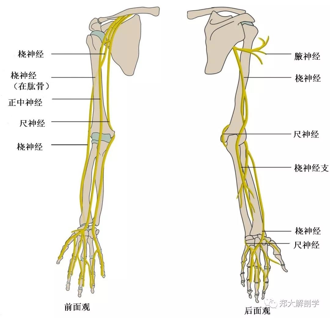
在上臂远端外侧,分开肱肌与肱桡肌就可以发现位于两者之间的桡神经.
图片尺寸1080x1194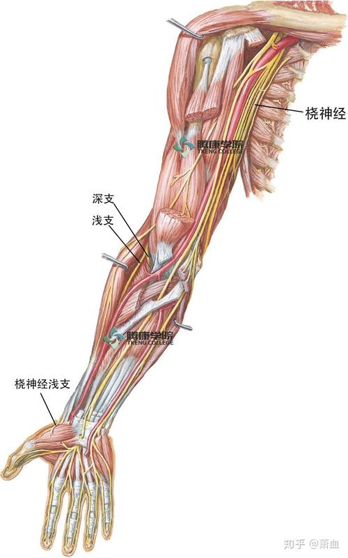
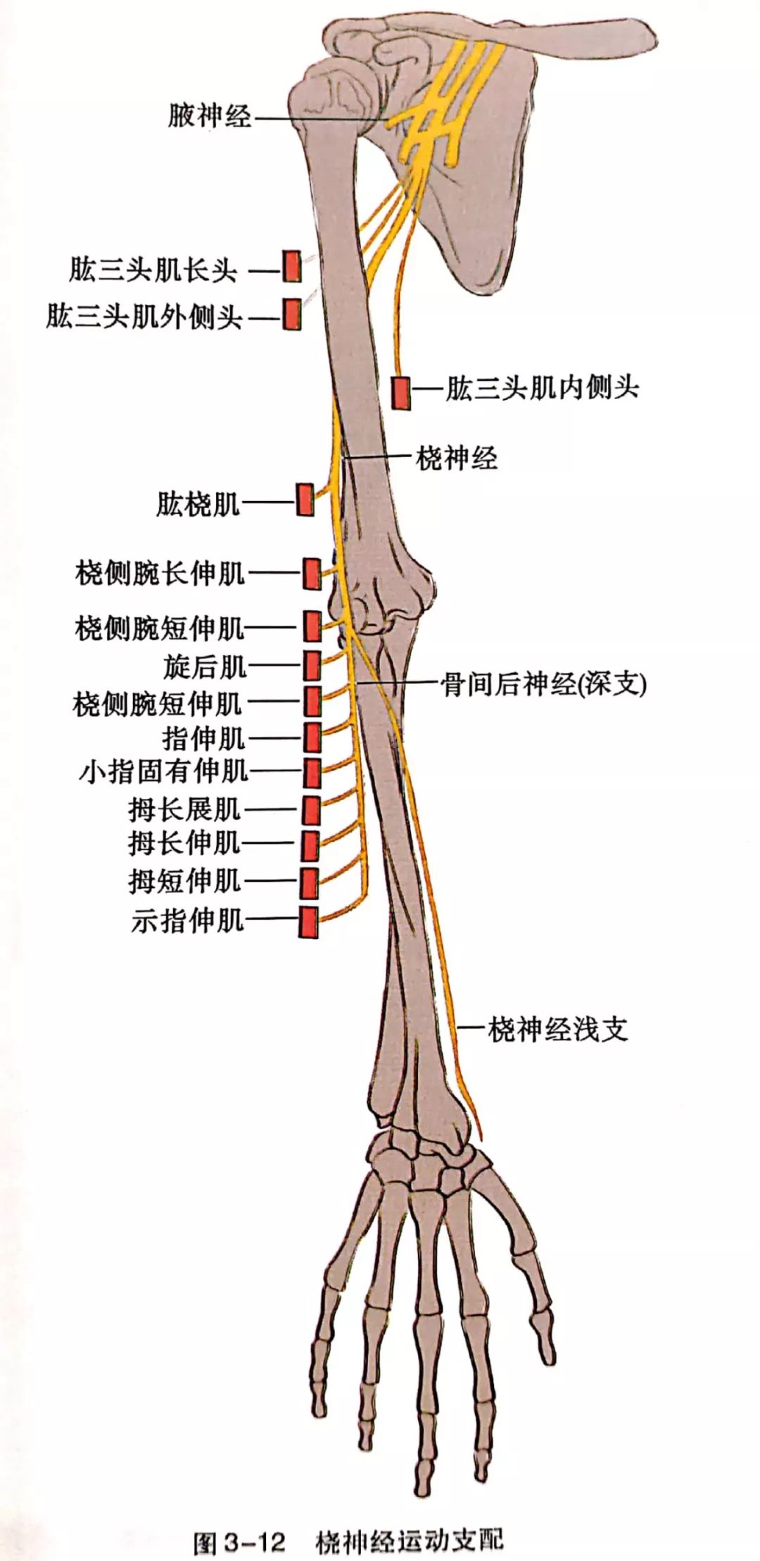
桡神经应用解剖
图片尺寸781x1250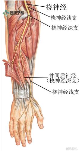
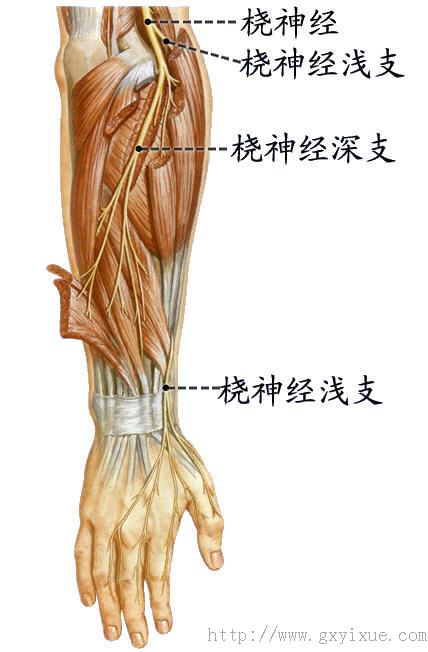
在位于肱骨外上髁和桡神经沟之间的一点,该神经发出它的两条终末支.
图片尺寸1080x2063
桡神经卡压的解剖学分析与查体及治疗定点
图片尺寸1080x2218
桡神经应用解剖
图片尺寸923x845
桡神经沟处损伤
图片尺寸640x796
大结节 b小结节 c外科颈 d解剖颈 e桡神经沟 2)关于
图片尺寸263x667
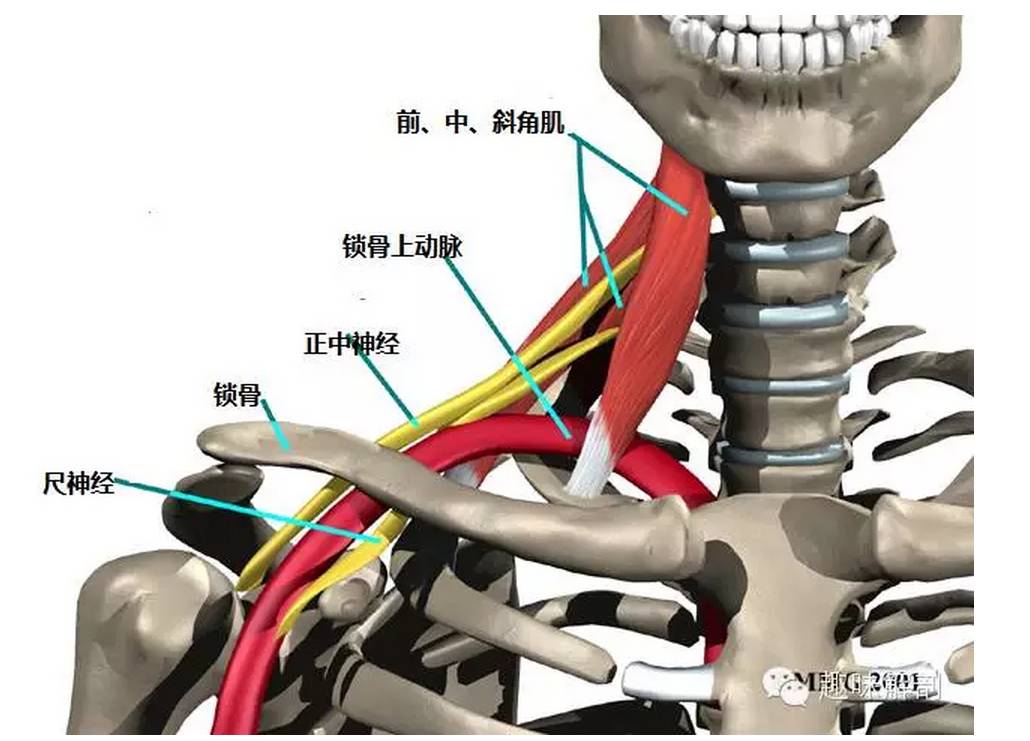
正中神经来源走行体表定位相关卡压
图片尺寸1080x1041
桡神经沟
图片尺寸268x287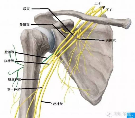
桡神经相关解剖和疾病
图片尺寸960x843
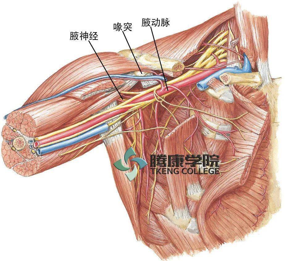
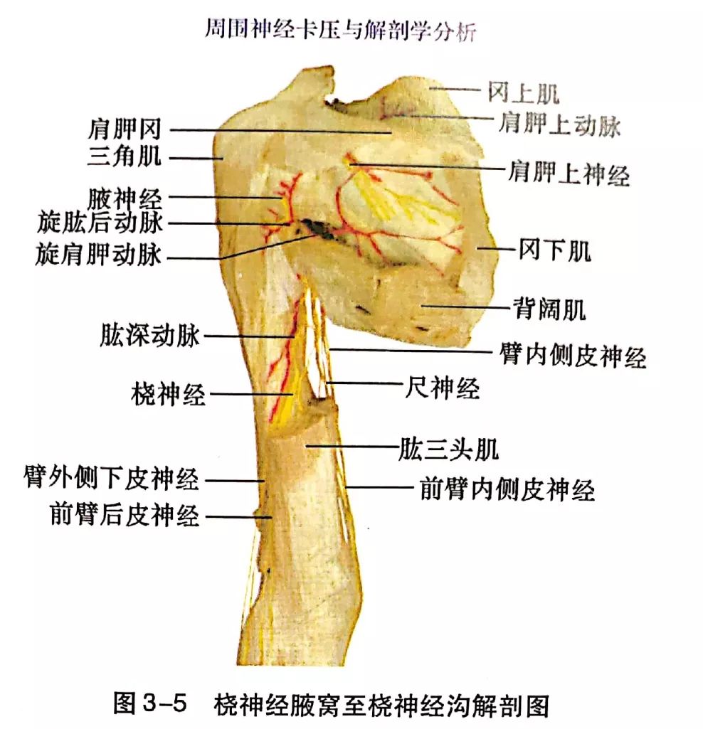
桡神经由c5~t1脊神经的神经纤维组成.该神经位于腋动脉的后下方.
图片尺寸1080x1342
(3)桡神经易被卡压的部位腋臂角处:在该部位桡神经正好位于肱骨颈和
图片尺寸1030x741
肱深动脉:伴随桡神经,在管 的中部分成前后2支(桡侧副
图片尺寸1080x810
腋窝至桡神经沟
图片尺寸988x1025
(3)桡神经易被卡压的部位腋臂角处:在该部位桡神经正好位于肱骨颈和
图片尺寸397x509