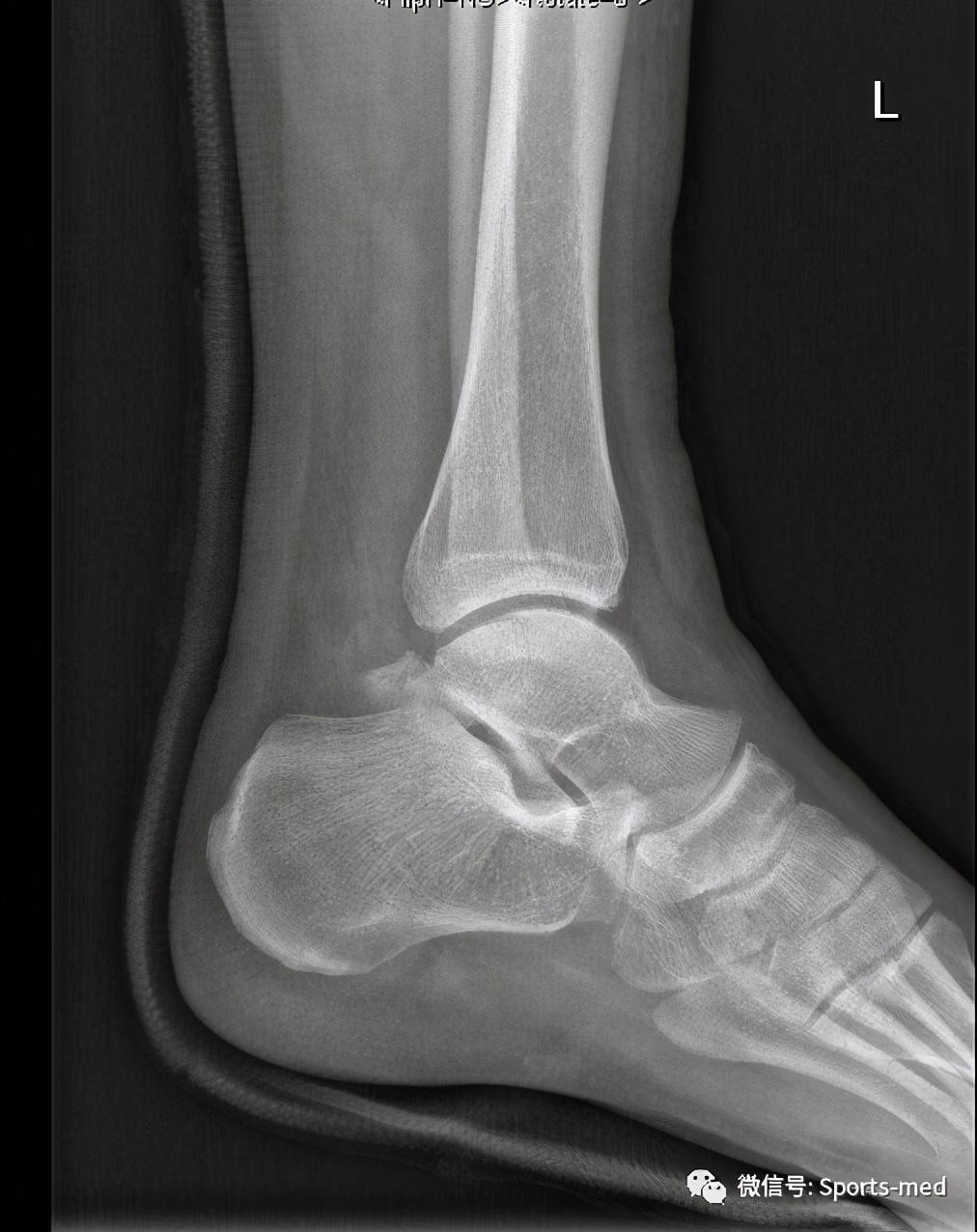
正常距骨x线图片

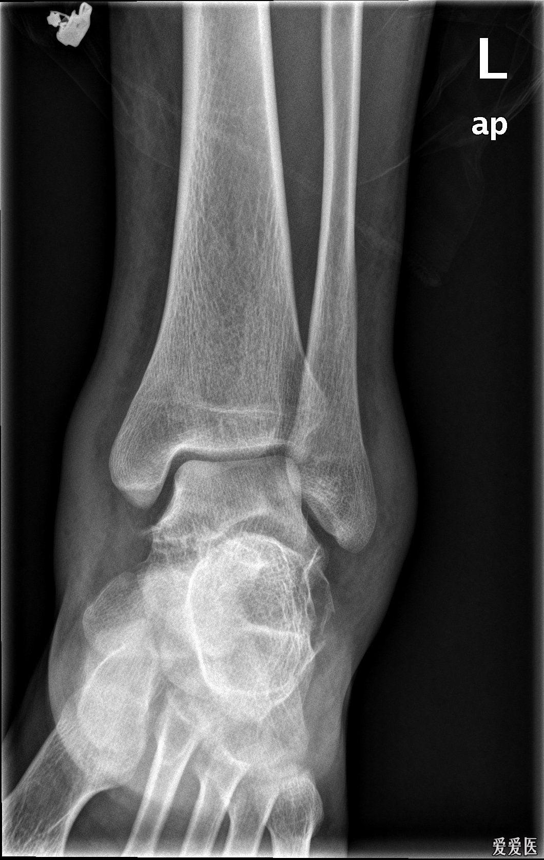
踝关节正位x线超声:可代替或结合mri检查软组织的损伤;ct:能进一步
图片尺寸632x720精品收藏距骨骨折和脱位放射科医生指南
图片尺寸437x500
距骨骨折关节镜治疗
图片尺寸1012x1276
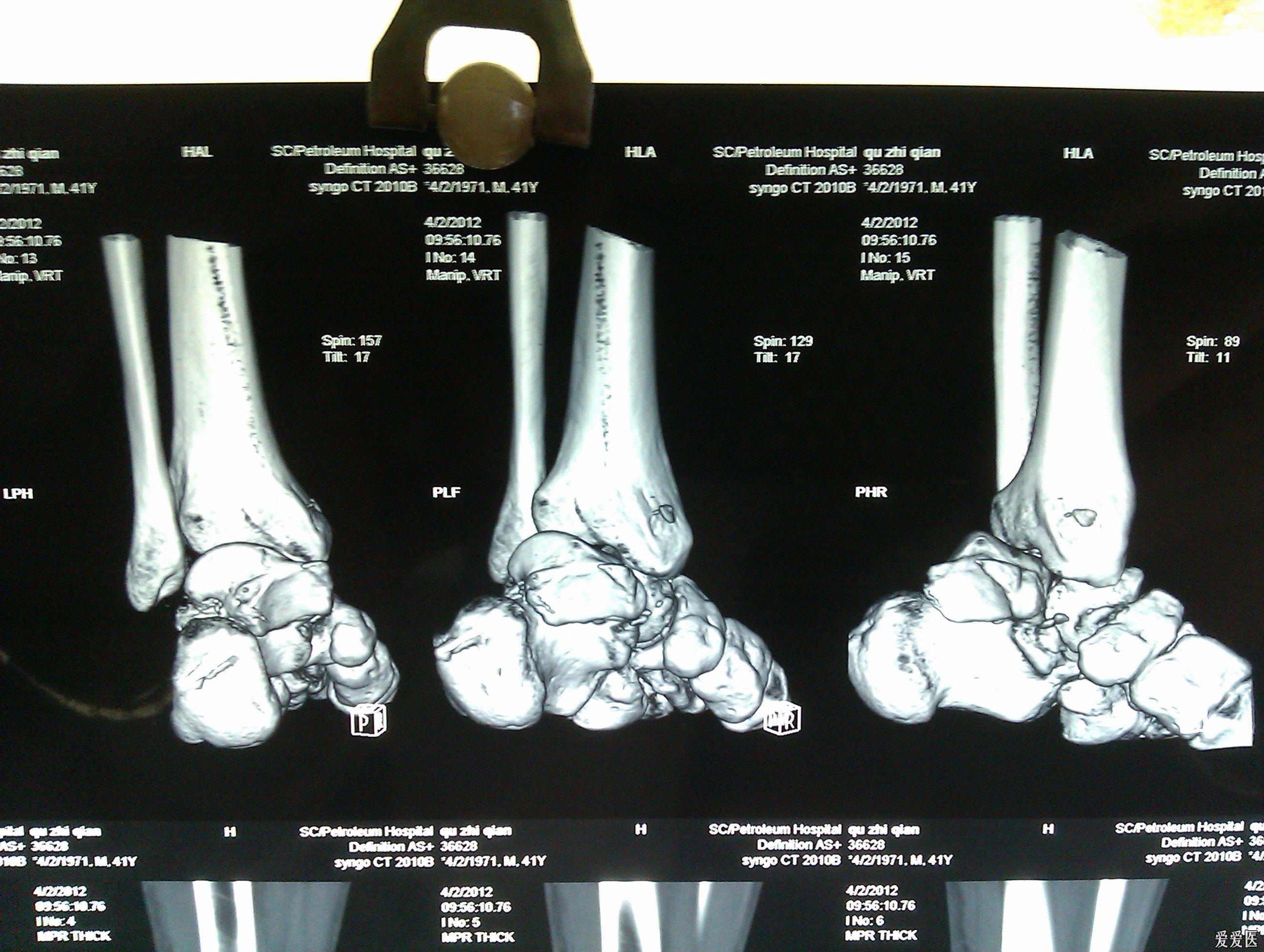
专业资源距骨骨折专题讲解
图片尺寸2592x1552
距骨骨折
图片尺寸680x907
左距骨骨折_1002_1001.jpg
图片尺寸1060x1675
距骨骨折
图片尺寸3256x2032
距骨骨折
图片尺寸680x907
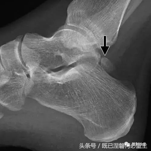
踝部的侧面x线片显示在距骨后方的椭圆形皮质骨化(箭头).
图片尺寸500x500
两例距骨体骨折病例—踝关节截骨入路螺钉内固定治疗距骨体骨折
图片尺寸1080x1080精品收藏距骨骨折和脱位放射科医生指南
图片尺寸499x500
两例距骨体骨折病例—踝关节截骨入路螺钉内固定治疗距骨体骨折
图片尺寸2000x2667
距骨骨折的诊断与治疗
图片尺寸2592x1952
距骨骨折脱位内外踝骨折急诊手术1例
图片尺寸3072x4096
距骨骨折
图片尺寸680x510
呈三角形, 与距骨后缘相切,三角骨与距骨后突外侧结节腓侧隐窝相对应.
图片尺寸410x558
腓骨下骨△ 儿童骨骺未闭△ 踝关节侧位△ 踝关节正位x线超声:可
图片尺寸640x669
5mm,骨折高度>7mm踝穴宽度 d线在距骨关节面下方5mm处,平 行于距骨
图片尺寸540x434
距骨骨折的诊断与治疗
图片尺寸2592x1952
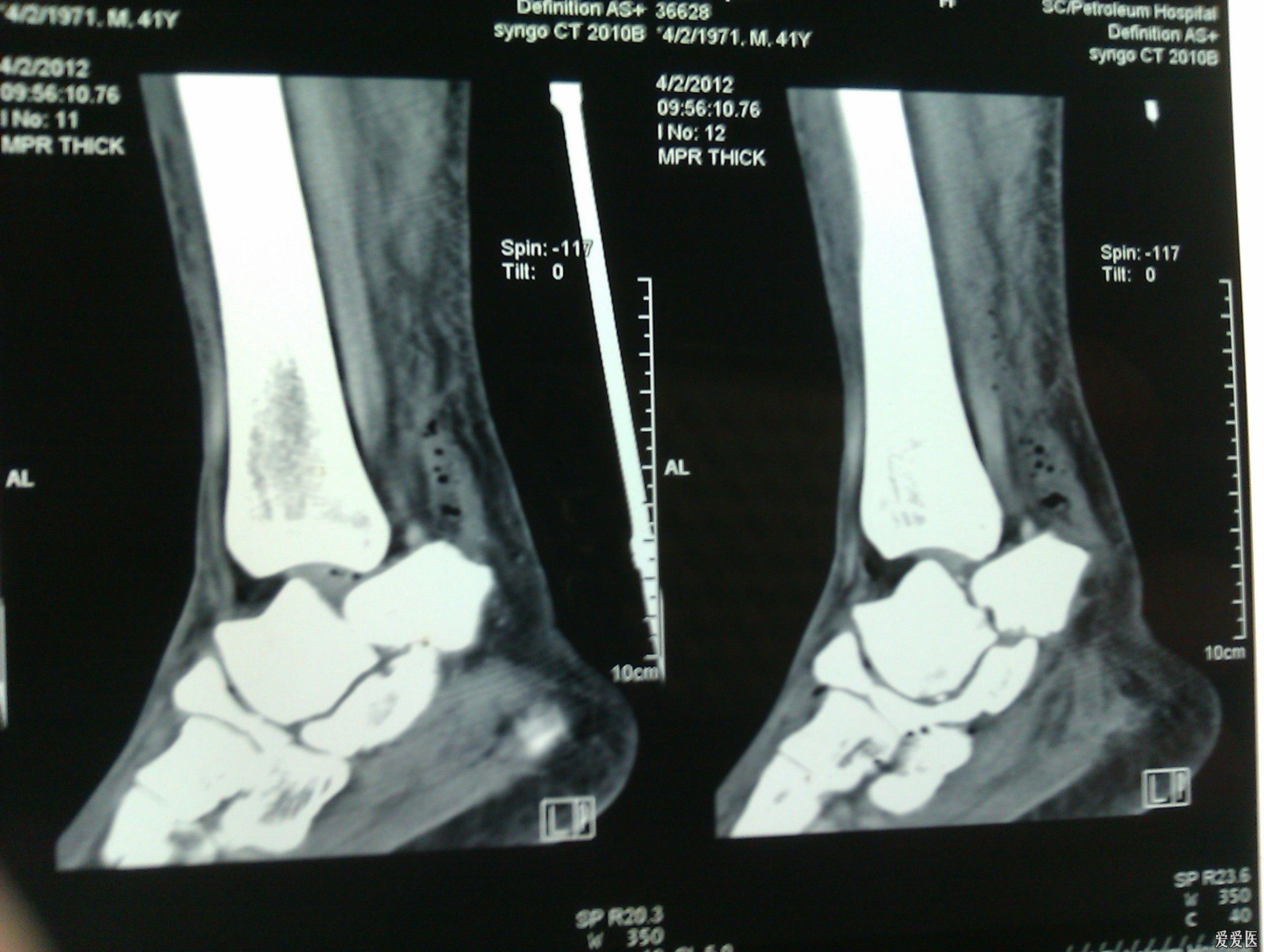
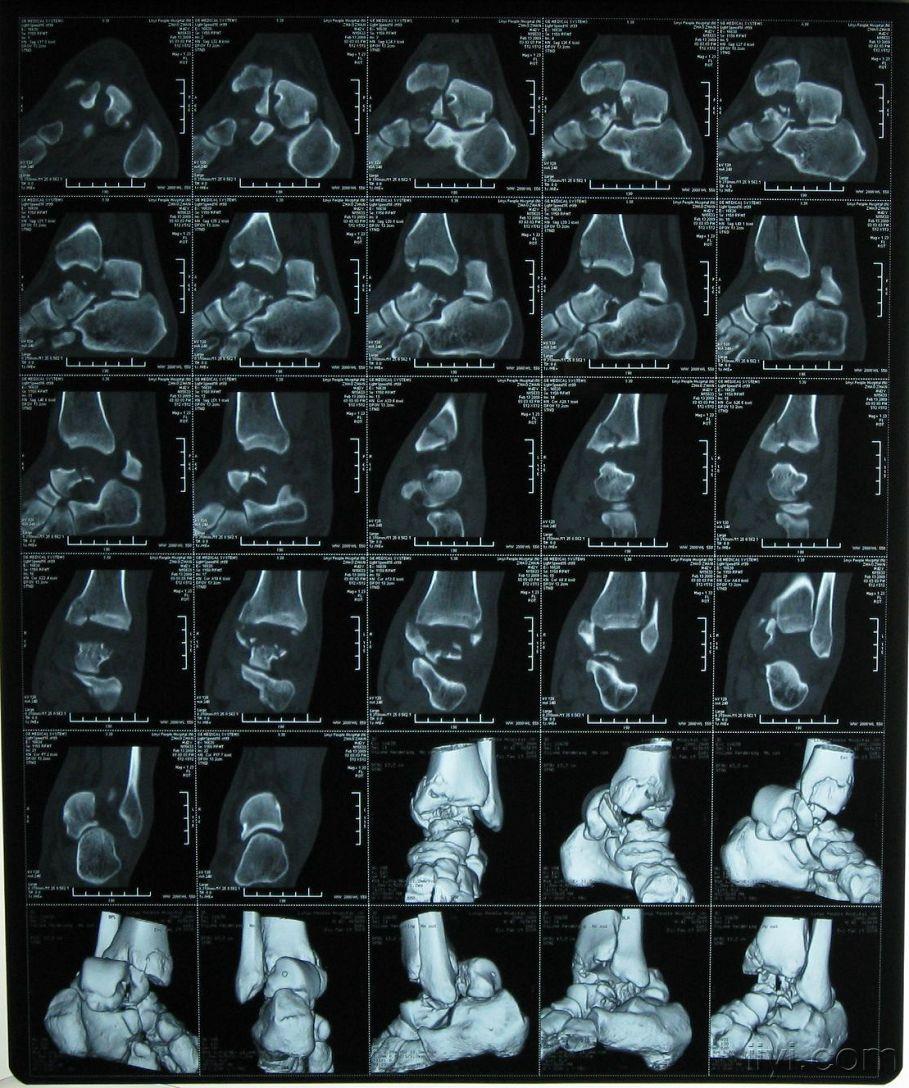
刚做的一例距骨骨折
图片尺寸909x1088


猜你喜欢:正常距骨ct图片距骨x线图片距骨侧位x线解剖图距骨3D图距骨正常图片距骨解剖图x线正常鼻骨x线图片正常足x线图片正位鼻骨骨折x线图片正常骶尾骨x线图片骶尾骨骨折x线图片正常足部x线图片正常足x线图片解剖图正常跟骨x线解剖图片正常足x线图片足正侧位x线正常图片正常膝关节x线图片正常肩关节x线图片正常鼻骨侧位x线图片正常的膝关节x片图片正常人的膝盖x片图片足斜位x线正常图片鼻骨x线图片侧面鼻骨侧位x线图片踝关节x线图片正常肩关节正位x片图正常脚跟骨x光片胃的正常x线像图片正常腹部x线平片图正常人尾骨图片舒筋枝的图片法国金丝雀干红葡萄酒黑白魔女库伊拉东方明珠素描简笔画三个人闺蜜头像 分开北京万城华府别墅颜辉陈望道老家扁桃体有白色东西是癌非诚勿扰贾明玉简介攻壳机动队2.0工地防护棚标准图片