气管隆突图片

如图气管隆突的高度相当于
图片尺寸305x336
沈阳市胸科医院完成东北首例ecmo下气管隆突重建手术
图片尺寸624x276
组成 : 气管软骨 ,平滑肌和结缔组织.
图片尺寸1080x810
(图2-1)2021年5月18日,全麻状态下,从喉罩插管顺利,声带正常,隆突锐利
图片尺寸714x366
5—2cm 长度11—13cm 第5-6胸椎水平分支 气管分叉角(隆突角) 60—85
图片尺寸1080x810
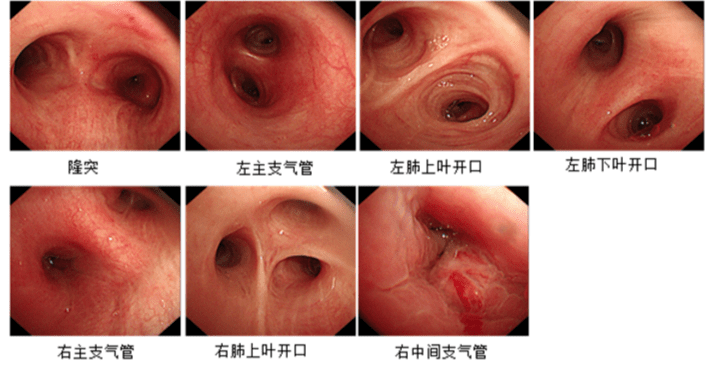
支气管镜图谱ppt课件ppt
图片尺寸800x600
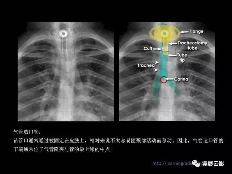
et管下端位于气管隆突上5—7cm这两幅图都是少量气腹.前10易漏诊.
图片尺寸750x563
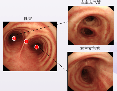
颈部基础 纤维内窥镜下的 气管腔和隆突上面观 气管腔下段 隆突 气管
图片尺寸1080x810
气管隆嵴
图片尺寸268x231
气管插管下隆突重度狭窄支架治疗
图片尺寸2000x2667
隆突重建气管成形术,解决特殊位置气管肿瘤难题
图片尺寸687x452
我看到如此镜下图像,我再三确认,镜子是在主气道内,确实是气管隆突
图片尺寸640x640
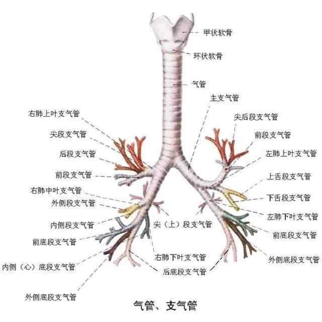
气管支气管解剖ppt
图片尺寸1080x810
气管隆突处观.jpg
图片尺寸640x680
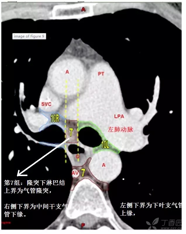
第7组:隆突下淋巴结 上界为气管隆突,左侧下界为下叶支气管上缘,右侧
图片尺寸636x800
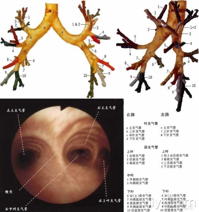
左主支气管与气管夹 角较大 右肺上叶支气管开口 距离气管隆突较近
图片尺寸1080x810
气管隆突位于气管的最下端,是左右支气管的分界部位.
图片尺寸380x295
气管四级三,各级支气管图 2 :左右主支气管气管隆突位于气管的最下端
图片尺寸336x330
纤支镜图谱ppt【最经典纤维支气管镜图谱】
图片尺寸1080x810
支气管 软骨环12~20个,多为15~16个 起于环状软骨(第6颈椎) 终于隆突
图片尺寸1080x810