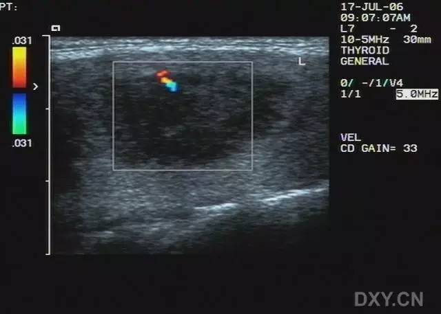
甲状腺上动脉超声图像

甲状腺结节的超声图像(图片来源:elizabeth cottril from thomas
图片尺寸684x410
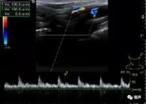
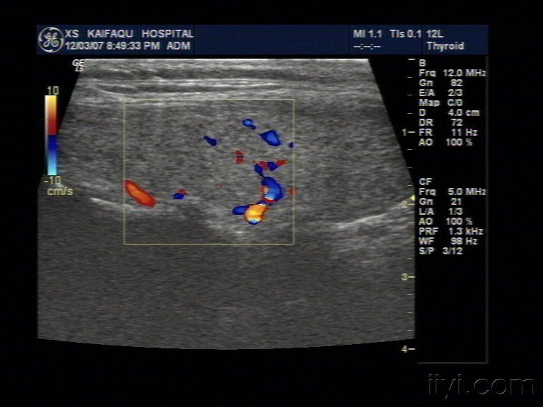
图 f:ht 患者,甲状腺上动脉收缩期峰值流速约 20 cm/s.
图片尺寸510x363
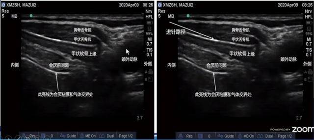
附着点(6)喉上神经内侧支(7)环状软骨(8)喉上动脉(9)甲状腺上动脉(10)
图片尺寸640x285
甲状腺超声诊断
图片尺寸1080x810
肿块血流较丰富,呈点条状及环绕血流信号,并可观察到有甲状腺下动静脉
图片尺寸1599x1199
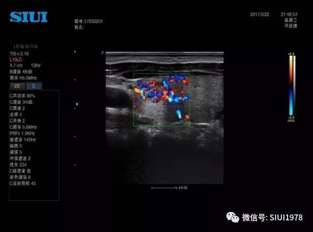
甲状腺多普勒超声图像可显示血管增生的复杂结节
图片尺寸640x435
体检超声发现甲状腺左叶实性肿物伴钙化,遂就诊.
图片尺寸600x218
甲状腺超声诊断
图片尺寸960x720
桥本甲状腺炎 - 超声医学讨论版 -丁香园论坛
图片尺寸4032x3016
这种甲状腺应该怎么诊断 - 超声医学讨论版 - 爱爱医
图片尺寸768x576
甲状腺功能亢进的超声表现
图片尺寸583x320
甲状腺超声报告怎么看?
图片尺寸640x490
【医海拾贝】甲状腺疾病的超声检查
图片尺寸690x517
这些甲状腺超声诊断误区不要犯
图片尺寸640x475
超声4c级,考虑ptc 飞鹰行动 07:58 病例二,上纵隔甲状腺病变,病灶与
图片尺寸600x800
扫查颈部血管时看到,患者去年做过甲状腺检查没有问题,该包块位于
图片尺寸768x576
超声典型病例:亚急性甲状腺炎
图片尺寸640x457
左甲状腺乳头状腺癌 手术病理证实 - 超声医学讨论版
图片尺寸768x576
典型的graves病甲状腺图片 - 超声医学讨论版 - 爱爱
图片尺寸768x576
于 2010-6-3 22:20 编辑 典型的超声图像主要表现为双侧甲状腺弥漫
图片尺寸800x924