疟原虫配子体图片

疟原虫镜检技术2013.3.ppt
图片尺寸1080x810
疟疾(malaria)是因疟原虫寄生人体组织而引起的寄生原虫病.
图片尺寸490x690
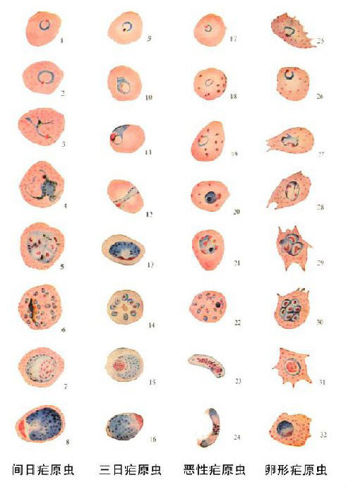
四种疟原虫镜下图(1)ppt
图片尺寸1080x810
▼(红)卵形疟原虫环状体(黑)卵形疟原虫雌配子体▼卵形疟原虫雄配子体
图片尺寸1080x1077
间日疟原虫#显微镜 #实验室 - 抖音
图片尺寸1440x1920
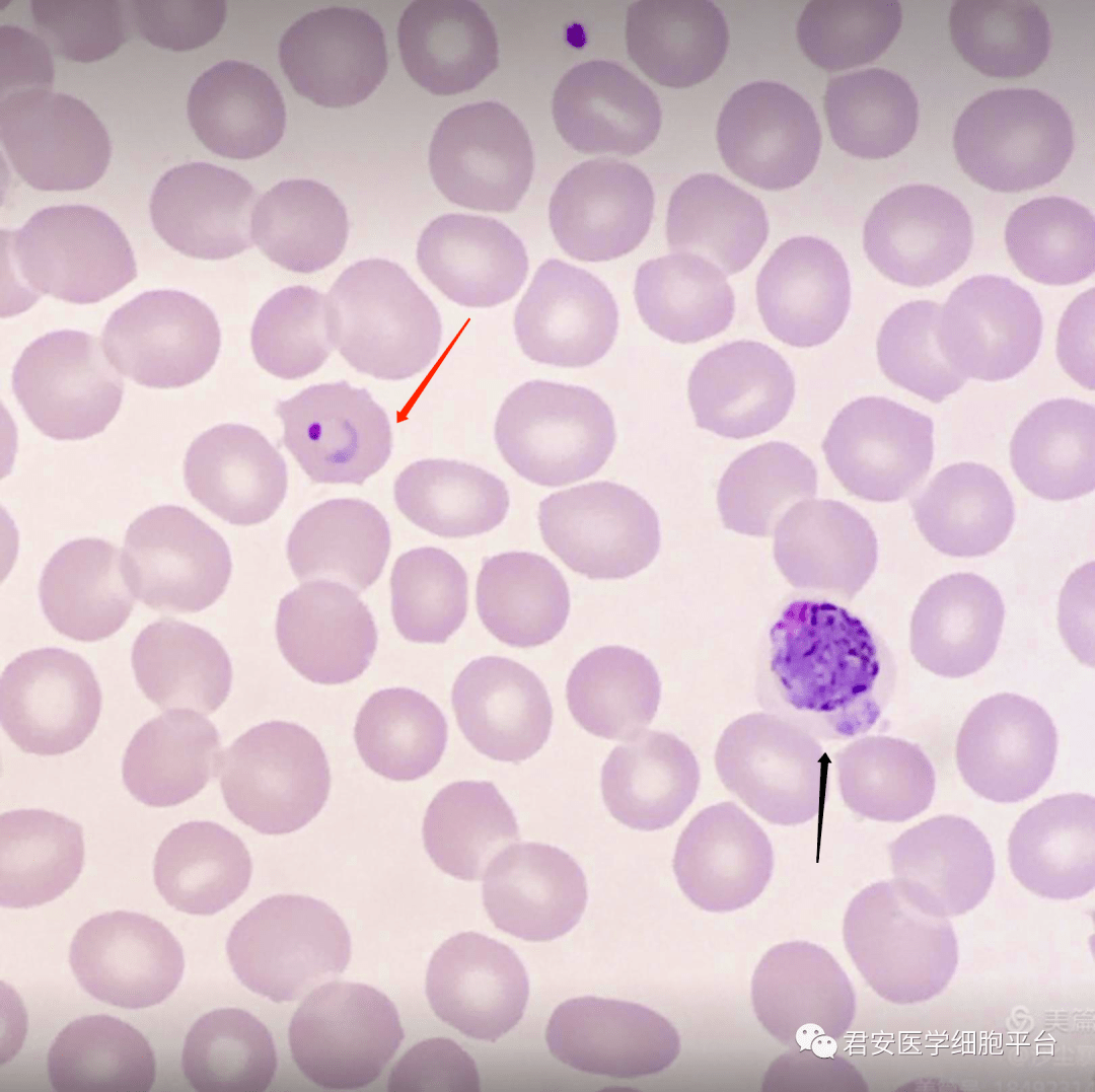
疟原虫环状体根据感染的疟原虫种类不同,疟疾可分为间日疟,恶性疟
图片尺寸1000x635
间日疟原虫雄配子体▼(黑)间日疟原虫雌配子体(红)间日疟原虫雄配子体
图片尺寸1080x1081
四种疟原虫镜下图(1)ppt
图片尺寸1080x810
绝对干货分享疟原虫检测技术
图片尺寸960x720
疟原虫镜检技术2013.3.ppt
图片尺寸1080x810
间日疟原虫配子体(雌)
图片尺寸1080x810
雌配子体:虫体大,占满整个红细胞,胞质深蓝;核小致密,深红色,位于虫体
图片尺寸2667x2000
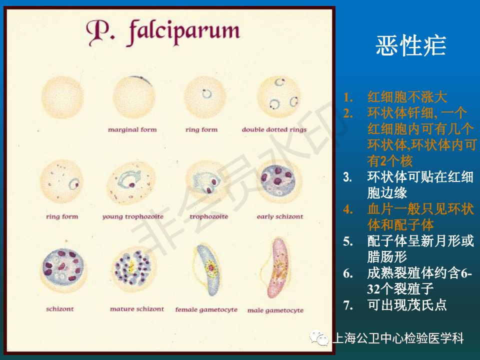
图中蓝色显示恶性疟原虫有性阶段-----配子体,红色圆形为红细胞.
图片尺寸600x599
三日疟原虫镜下形态
图片尺寸1080x810
卵形疟原虫镜下形态
图片尺寸1620x2160
三日疟原虫镜下形态
图片尺寸1620x2160
裂殖体(schizont) 滋养体成熟后,疟原虫的细胞核开始不断的分裂.
图片尺寸140x140
常见疟原虫血片
图片尺寸1080x1439
疟原虫的镜检技术-2014.05ppt
图片尺寸1080x810
四种疟原虫镜下图(1)ppt
图片尺寸1080x810