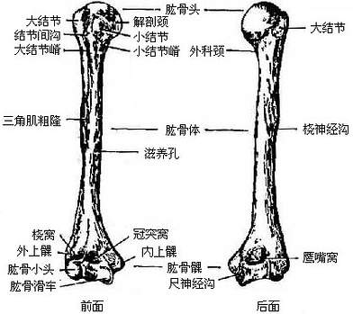
肱骨大结节嵴

骨科医生必备!肱骨大结节骨折的诊断与治疗
图片尺寸952x734
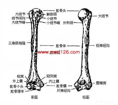
人体肱骨解剖示意图-人体解剖图
图片尺寸694x693
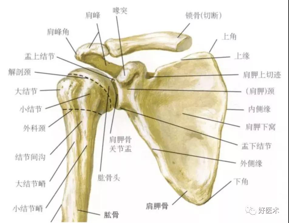
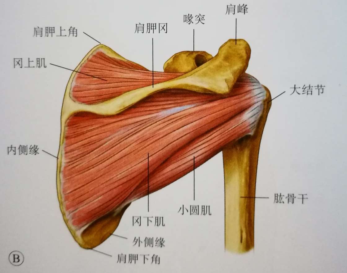
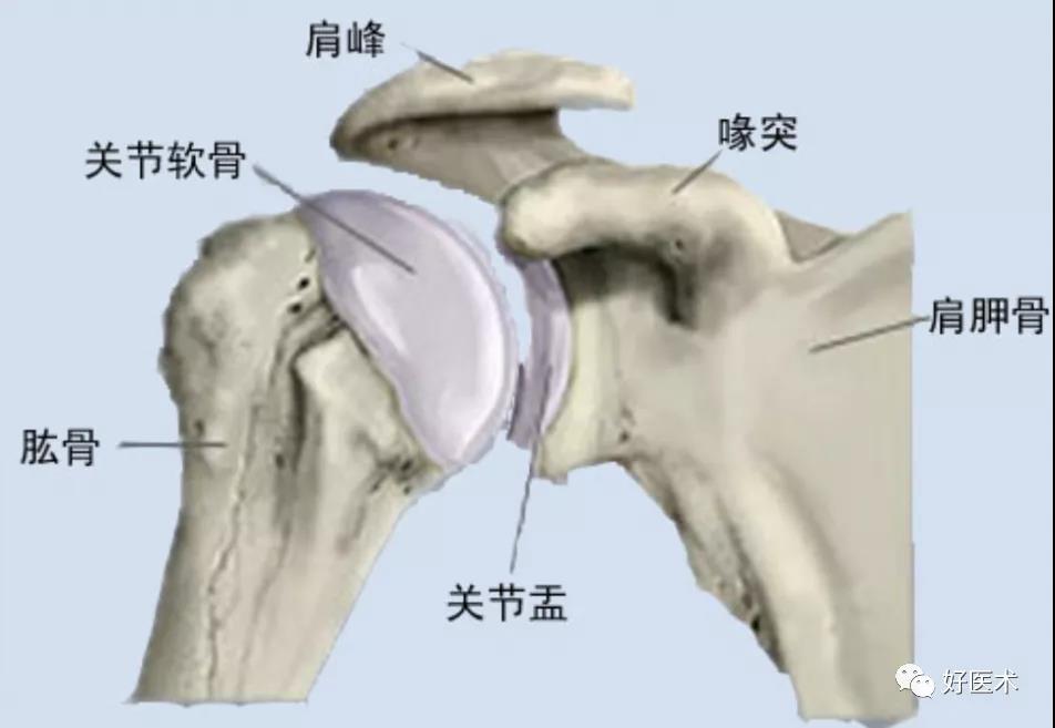
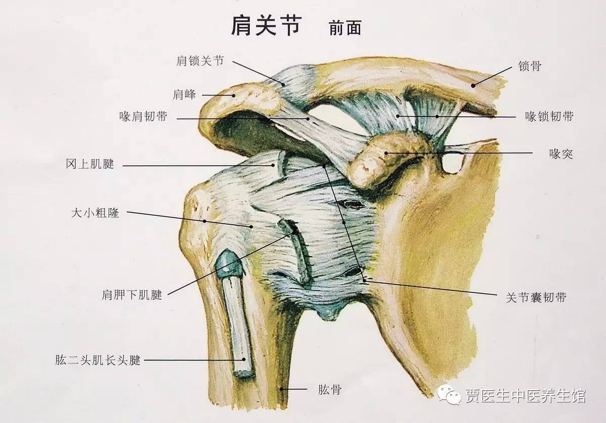
肩部: 肩峰 肱骨大结节 肩胛冈 锁骨 喙突 2.
图片尺寸1080x810
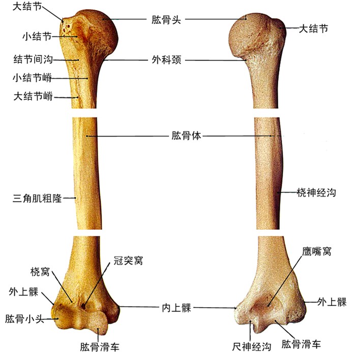
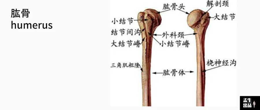
肱骨
图片尺寸1080x810
喙肩韧带及肩峰下滑囊下面,肩关节囊上面,止于肱骨大结节上切迹,其
图片尺寸967x1228
上端膨大,向内上方突出的半球形的关节面,叫做肱骨头(图3-19),与肩胛
图片尺寸392x349
四肢骨骼解剖和骨折ppt
图片尺寸1080x810
肱骨头的外侧和前方有隆起的大结节和小结节,向下各延伸一嵴,称大结节
图片尺寸660x873
图2-10 肩胛骨(1)肱骨:为长骨,分为上,下两端和肱骨体.
图片尺寸486x472
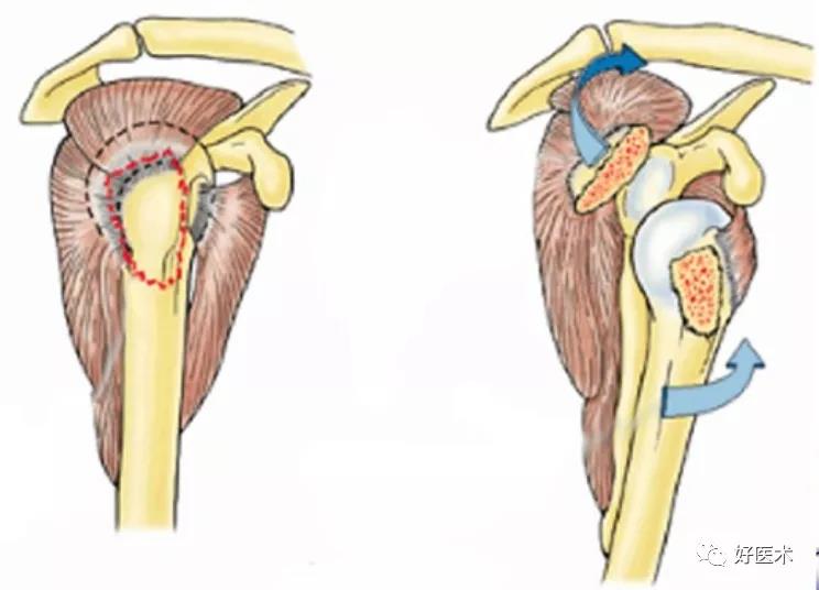
肱骨大结节骨折治疗
图片尺寸1120x879
骨科医生必备!肱骨大结节骨折的诊断与治疗
图片尺寸952x657
肱骨内,外上髁:位于上 臂下端内外两侧皮下最 为隆凸处.
图片尺寸1080x810
上端膨大,向内上方突出的半球形的关节面,叫做肱骨头(图3-19),与肩胛
图片尺寸449x400
小结节下方的为小结节嵴大结节下方的为大结节嵴大结节与小结节是肱骨
图片尺寸900x383
肱骨大结节骨折
图片尺寸2250x2491
最新 骨科教学病例讨论(肱骨近端骨折)ppt
图片尺寸1080x810
「医学笔记」——肱骨近端骨折(一)|大结节|胸大肌|切口_网易订阅
图片尺寸640x422
骨科医生必备!肱骨大结节骨折的诊断与治疗
图片尺寸744x536
上端有肱骨头,上端的前外侧面为大结节,内侧为小结节.
图片尺寸1080x1439
2,肱骨小结节,结节嵴:位于肱骨头前内侧,是肩胛下肌,喙肱韧带的止点
图片尺寸1200x839