肾小管上皮细胞模式图

肾小管上皮细胞怎么检测
图片尺寸1055x820
小鼠肾小管上皮细胞
图片尺寸400x284
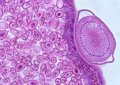
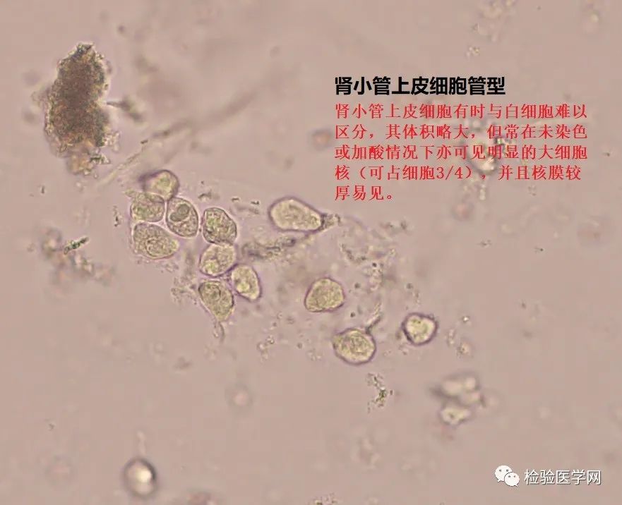
显微镜下为肾小管单纯性上皮细胞
图片尺寸900x1200
肾小管上皮细胞怎么检测
图片尺寸958x991
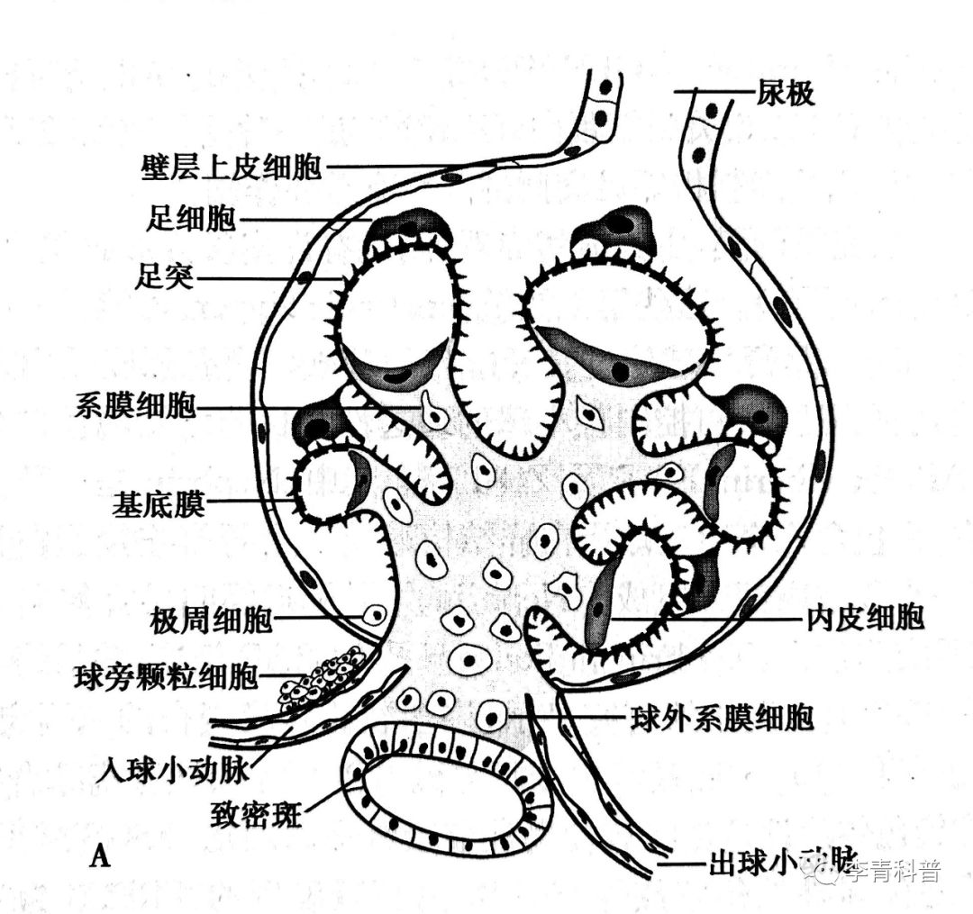
尿极 血管球 远曲小管 球旁细胞 近曲小管 血管极 第14页 (共21
图片尺寸1080x810
肾小管上皮细胞怎么检测
图片尺寸1585x565
如图为肾小管上皮细胞重吸收原尿中na
图片尺寸519x231
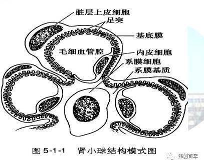
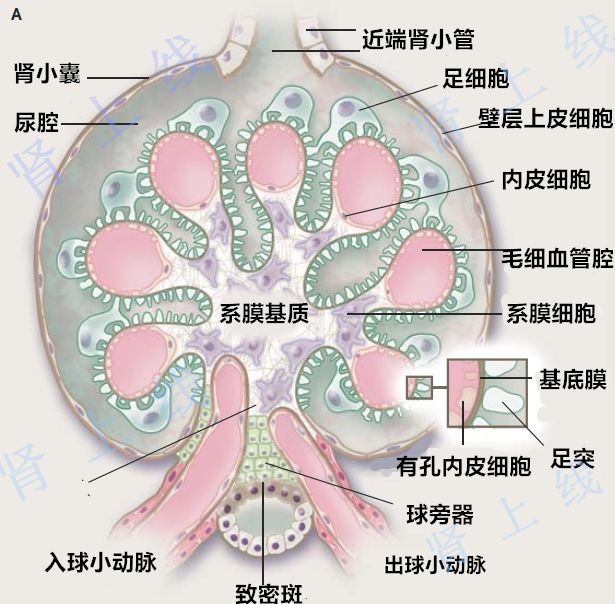
肾小球毛细血管壁由内皮细胞,基底膜,脏层上皮细胞(足细胞)构成,形成
图片尺寸407x330
上皮细胞形态图谱 肾小管上皮细胞 肾小管上皮细胞
图片尺寸1080x810
肾小管ppt
图片尺寸1080x810
管-球反馈 | sglt2 抑制剂护肾的主要机制_细胞
图片尺寸1080x1015
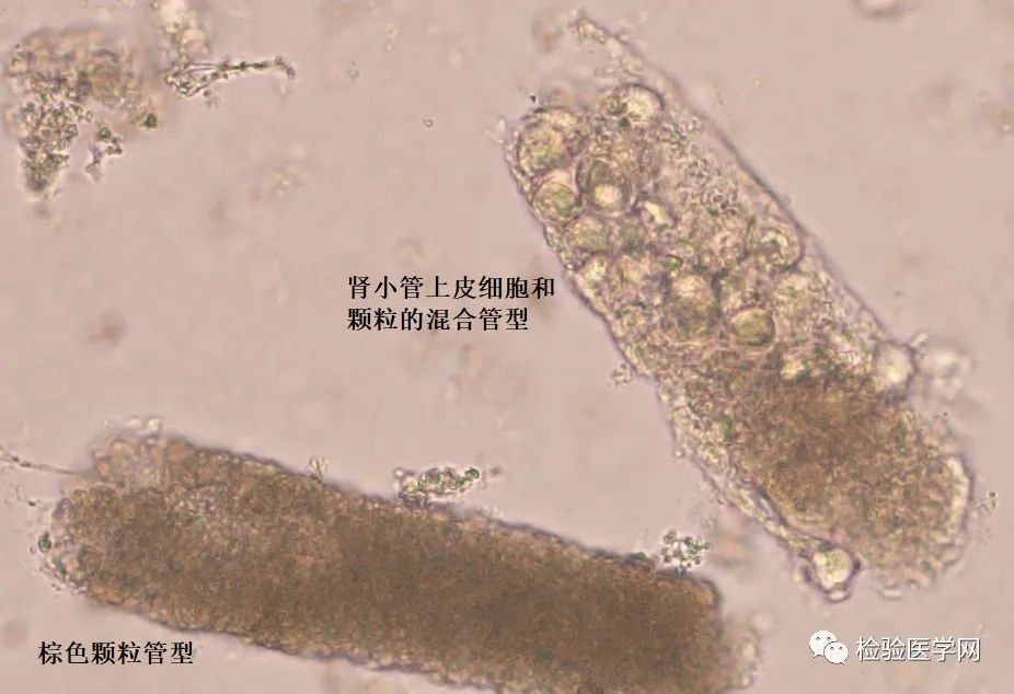
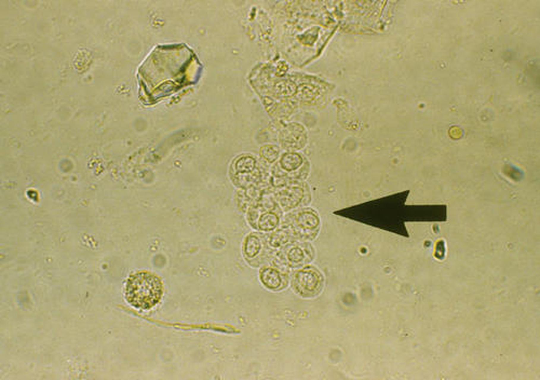
回到本例标本,其中红细胞少见,并可发现较多脱落的肾小管上皮细胞及
图片尺寸926x634
泌尿系统完整结构ppt
图片尺寸1080x810
科普知识竞赛#尿中所见上皮细胞由肾小管,肾盂,输尿管,膀胱,尿道荡Ζ
图片尺寸1818x1280
葡萄糖在肾小管上皮细胞易化扩散
图片尺寸1451x1164
病理实验绘图
图片尺寸1080x691
研究发现,在大多数膜性肾病患者体内能够检出抗肾小球足细胞上的磷脂
图片尺寸615x604
回到本例标本,其中红细胞少见,并可发现较多脱落的肾小管上皮细胞及
图片尺寸881x716
肾小体 肾小囊 肾小球 肾小管 毛细血管 集合管
图片尺寸1080x810
肾小球血管袢薄而清 晰.
图片尺寸1080x810