胸膜CT

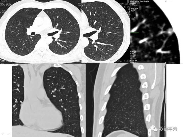
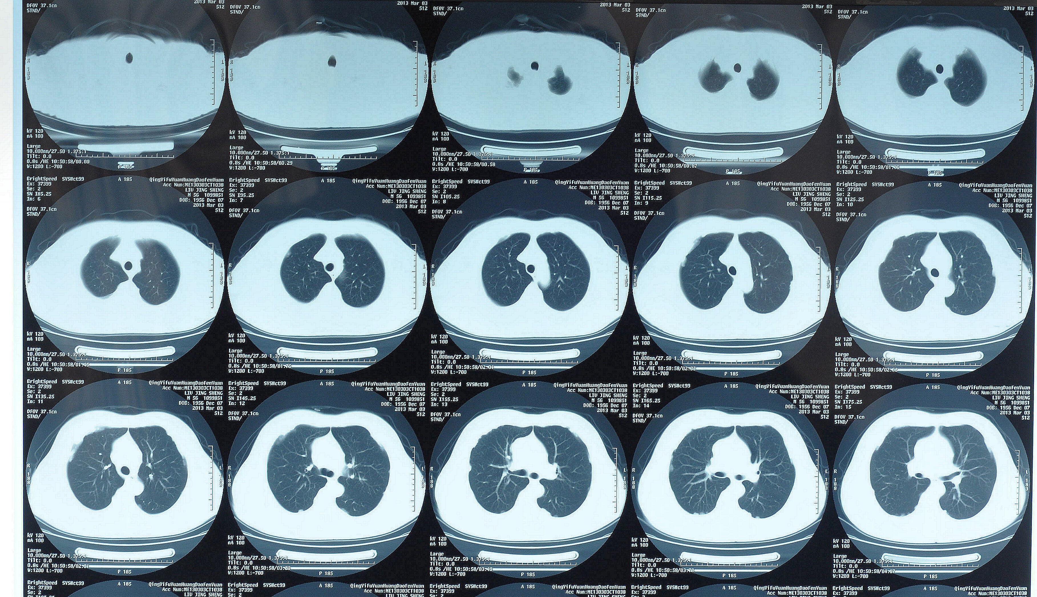
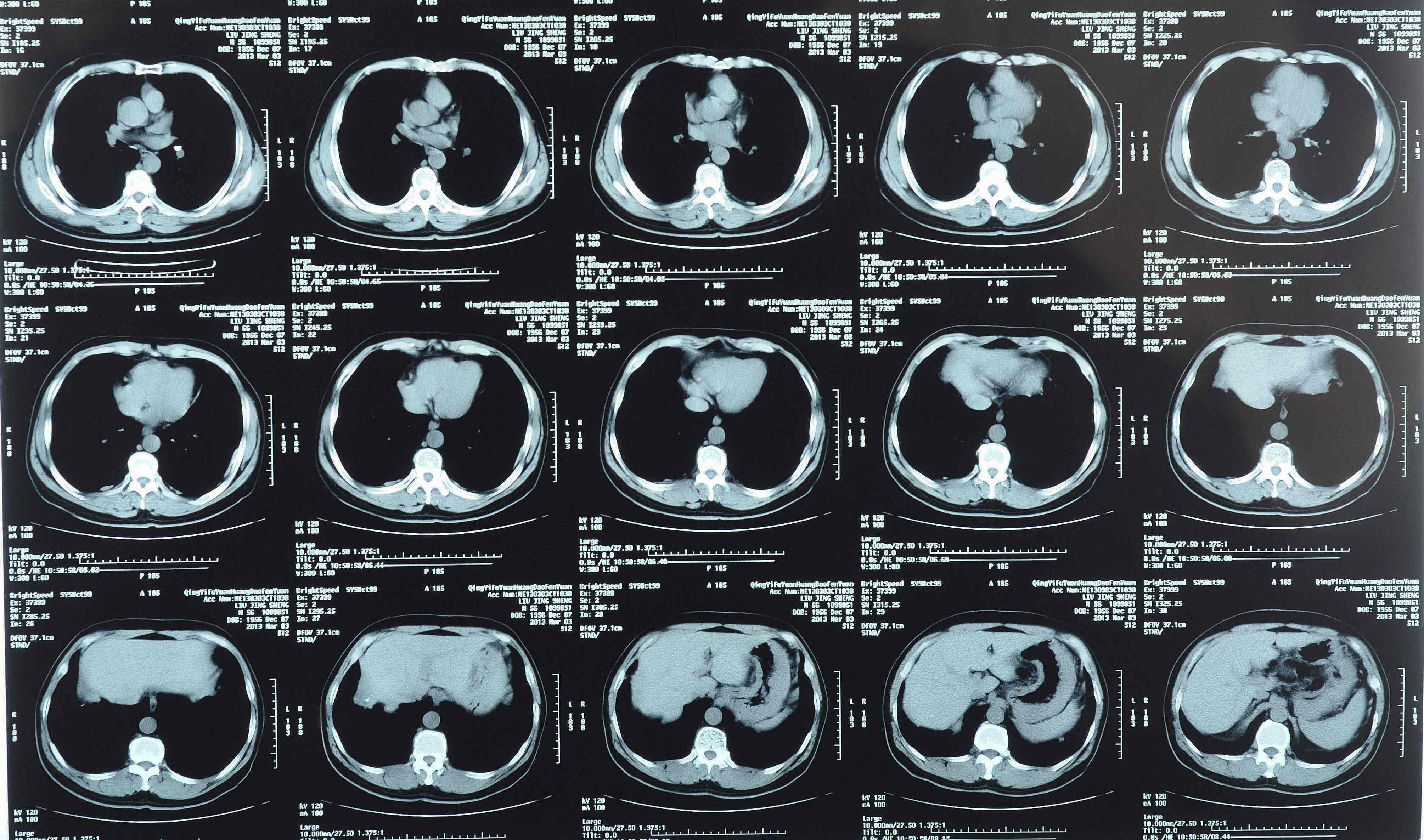
ct右肺胸膜发现了什么?
图片尺寸3459x1728
ct胸膜下点条状的线提示什么
图片尺寸1617x1163![【病例讨论】结核性胸膜炎? [病例帖]](https://i.ecywang.com/upload/1/img2.baidu.com/it/u=273888009,3730541851&fm=253&fmt=auto&app=138&f=JPEG?w=516&h=483)
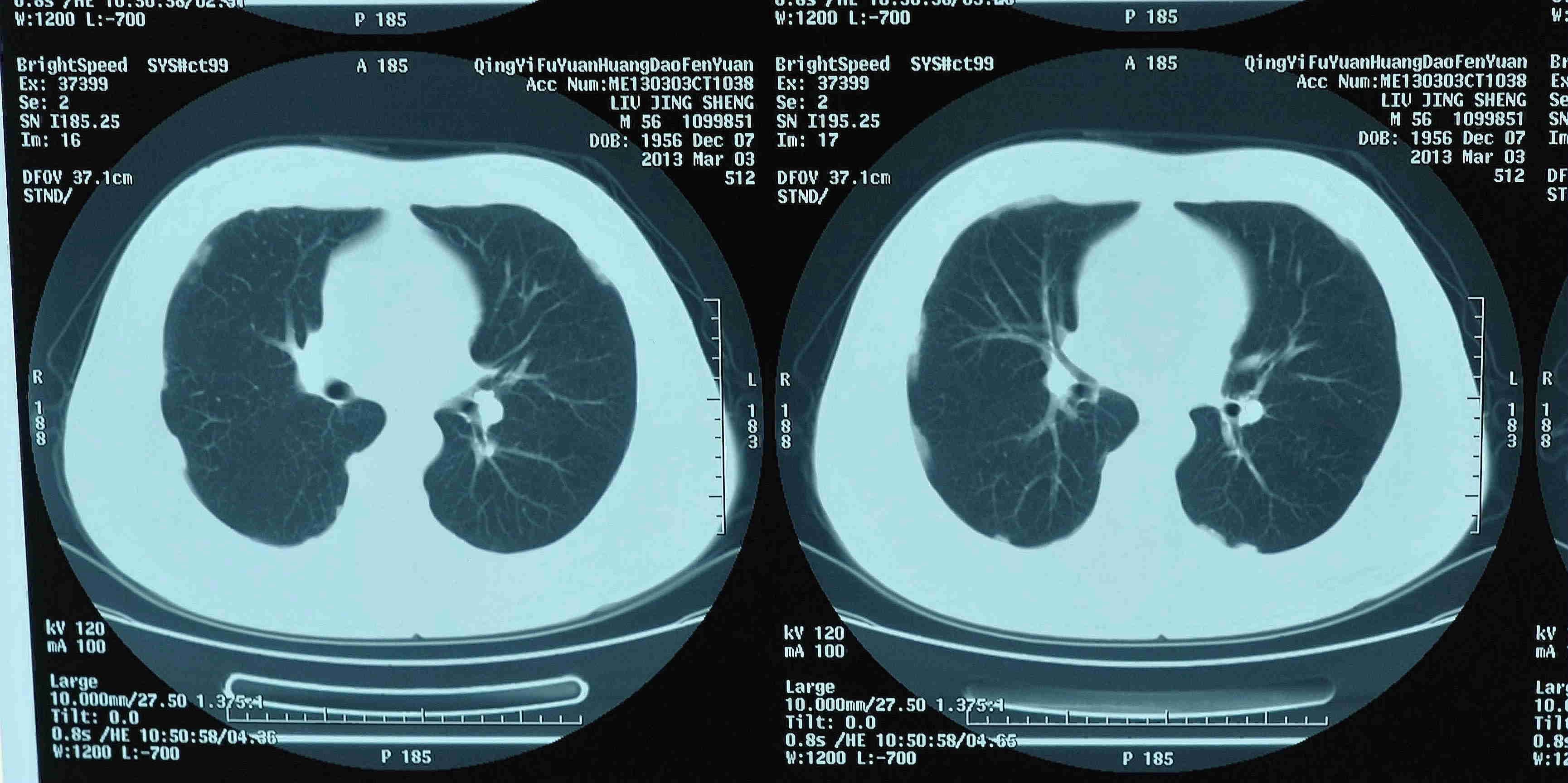
【病例讨论】结核性胸膜炎? [病例帖]
图片尺寸516x483
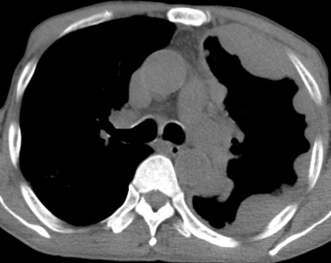
图2:胸部ct显示左侧胸膜增厚钙化.
图片尺寸515x449
ct征象对早期肺腺癌分型之胸膜凹陷征
图片尺寸640x480
影像知识科普胸膜增厚钙化在ct上什么表现专科医生讲解
图片尺寸1024x576
胸膜间皮瘤1例ct影像鉴别
图片尺寸609x1495
胸部ct学习系列之一胸膜病变
图片尺寸687x535
ct右肺胸膜发现了什么?
图片尺寸3782x2205
胸膜炎|引流术|ct|-健康界
图片尺寸640x959
ct右肺胸膜发现了什么?
图片尺寸3880x2130
ct右肺胸膜发现了什么?
图片尺寸3635x2093
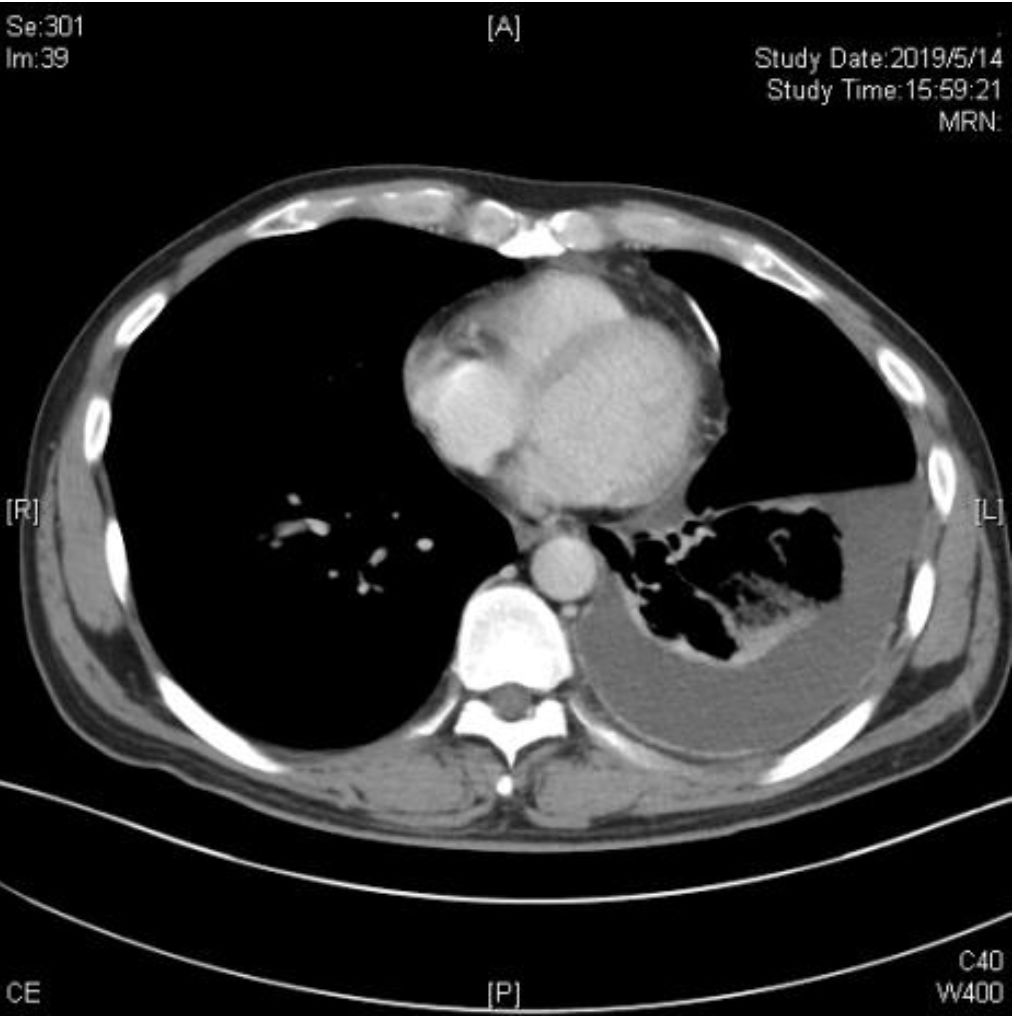
图2:胸部增强ct显示左侧胸腔积液及胸膜增厚
图片尺寸1014x1016
以下是典型恶性胸膜间皮瘤的ct表现mpm发病率呈上升趋势,预后差,在
图片尺寸672x534
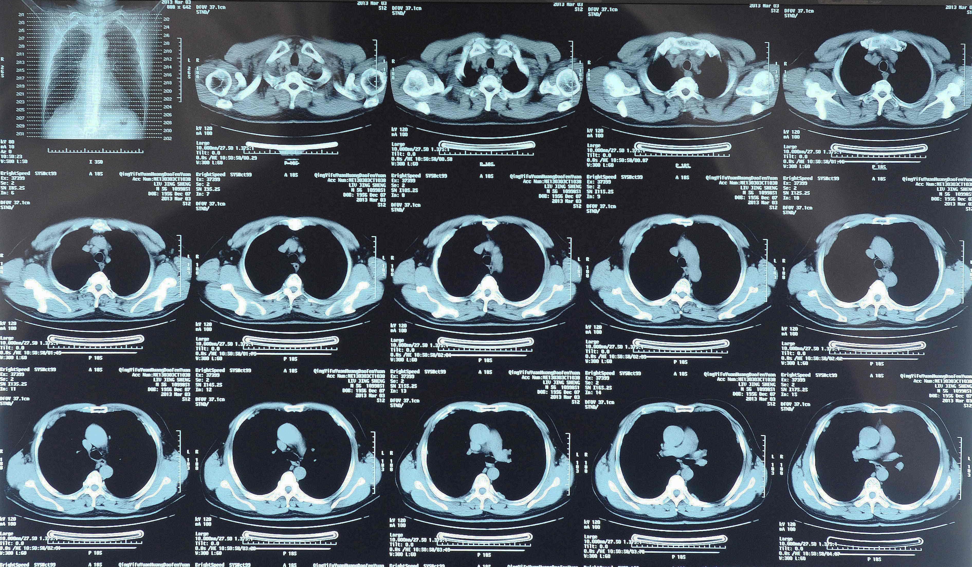
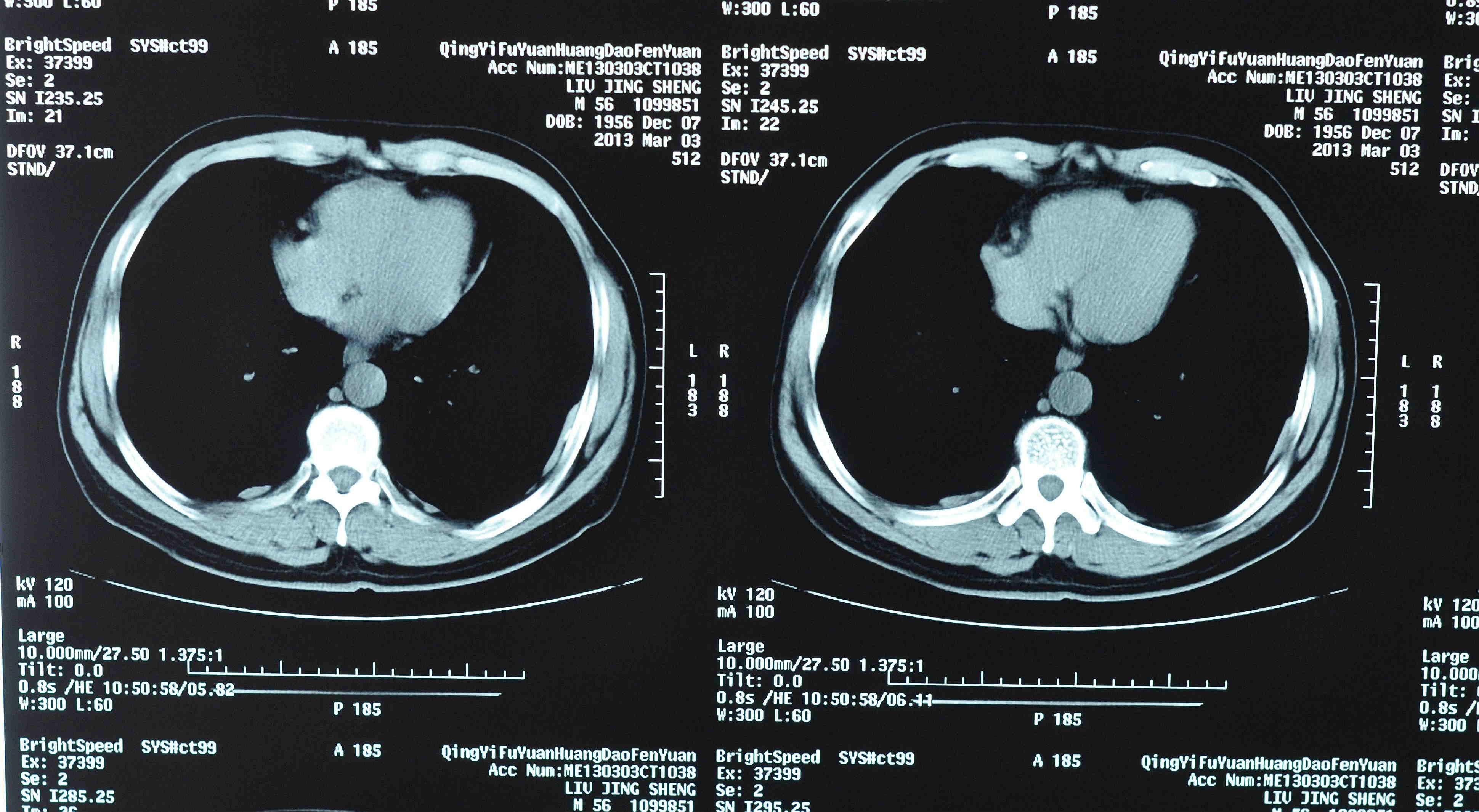
我年前得了结核性胸膜炎,已经治疗了7个月了,昨天去医院做ct诊断说有
图片尺寸2736x4864
ct右肺胸膜发现了什么?
图片尺寸4019x2372
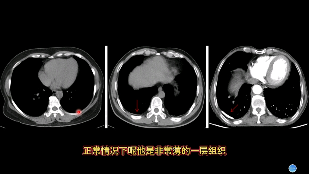
我们在阅胸片或ct的时候,很大一部分病人会合并有局部胸膜增厚
图片尺寸1000x988
我年前得了结核性胸膜炎,已经治疗了7个月了,昨天去医院做ct诊断说有
图片尺寸2736x4864
丁香园论坛
图片尺寸2448x2448
【讲座】病例分析推演:恶性胸膜间皮瘤的ct诊断和鉴别诊断
图片尺寸960x720

![【病例讨论】结核性胸膜炎? [病例帖]](https://img.dxycdn.com/upload/2013/07/08/59/34031104.snap.jpg)