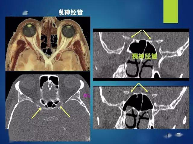
视神经孔ct解剖图

ct影像眼的附属结构:由眼外肌,视神经,泪器以及眶内的脂肪,血管,神经
图片尺寸851x397
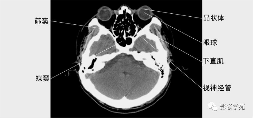
眼眶横断面(视神经层面)
图片尺寸1077x808
外伤骨折 鼻骨 眼眶 视神经管 骨折
图片尺寸1080x2160
外伤骨折 鼻骨 眼眶 视神经管 骨折
图片尺寸1080x2160
多层螺旋ct三维重建在视神经管影像解剖的应用ppt
图片尺寸1080x810
收藏最全的12对颅神经解剖及影像
图片尺寸640x480
多层螺旋ct三维重建在视神经管影像解剖的应用ppt
图片尺寸1080x810
上海交大医学院断层解剖学,考试复习必备! 视神经与视交叉冠状层面
图片尺寸1080x810
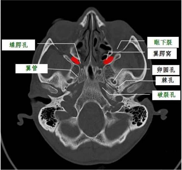
翼腭窝的ct影像解剖
图片尺寸632x590
多层螺旋ct三维重建在视神经管影像解剖的应用ppt
图片尺寸1080x810
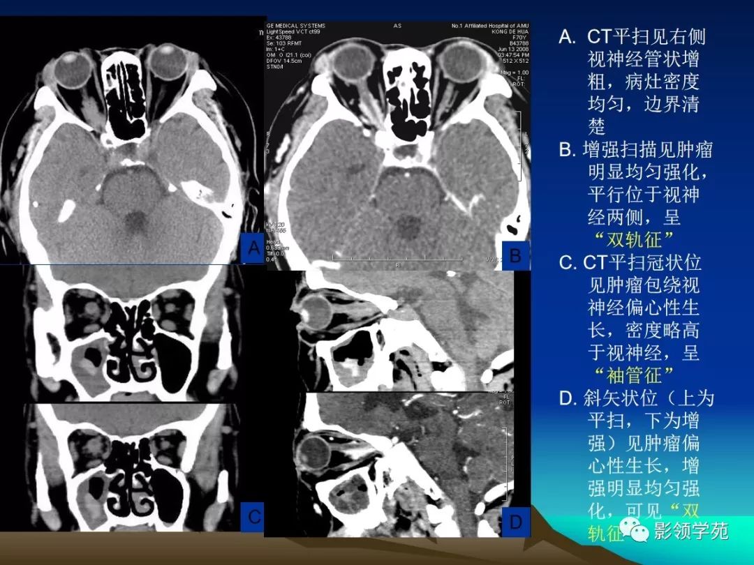
患者左眼睑青紫肿胀明显,视物模糊,经ct检查示:视神经管上壁骨折并
图片尺寸325x354
外伤骨折 鼻骨 眼眶 视神经管 骨折
图片尺寸1080x2160
正常眼眶ct解剖 1眼眶横断面(晶状体层面) 2眼眶横断面(视神经层) )
图片尺寸605x278
2,术后复查ct显示左眼视神经管外侧壁有效开放,vep诱导出有效波形手术
图片尺寸1601x798
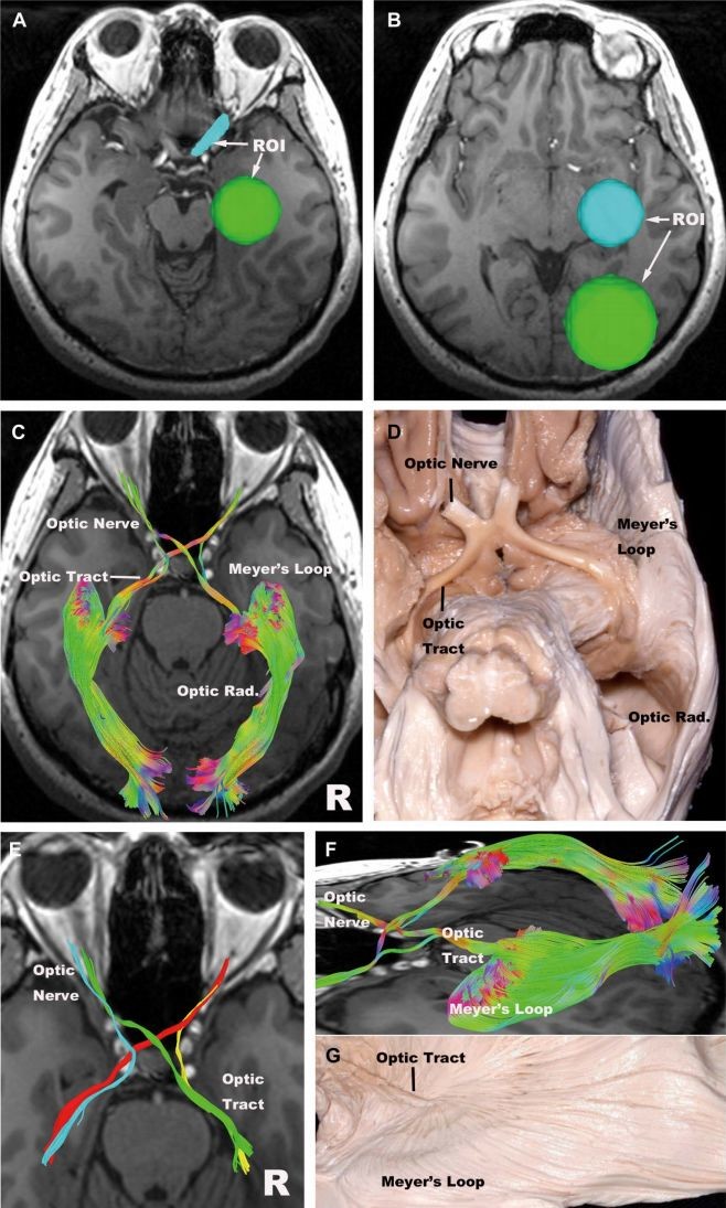
roi分别置于视神经和外侧膝状体;b.roi分别置于外侧膝状体和枕叶;c.
图片尺寸658x1095
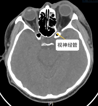
视神经管
图片尺寸1080x810
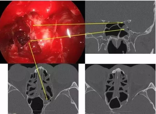
手术视频同仁医院康军内镜经鼻入路眶视神经管区病变的手术治疗要点
图片尺寸606x456
眼和眼眶的影像学诊断
图片尺寸1080x810
【ccos2016神经】梁申芝教授:人视神经高分辨mri及多平面重建技术的
图片尺寸1081x446
手术视频同仁医院康军内镜经鼻入路眶视神经管区病变的手术治疗要点
图片尺寸601x440


猜你喜欢:视神经管ct解剖图视神经ct断层解剖图视神经管ct解剖视神经管解剖图视神经ct解剖正常图像视神经分段解剖图视神经解剖结构图视神经解剖图视神经解剖视神经管骨折ct图像视神经图解视神经在脑部图视交叉ct断层解剖图视神经示意图视神经在头部位置图视神经分布图视神经位置图视神经图肺动脉解剖图ct图视神经管ct扫描范围头颅解剖图ct髋关节ct解剖图髋关节ct断层解剖图正常人的视神经图片上颌窦解剖图ct心脏ct横断面解剖图肺部ct解剖图小肠ct分段解剖图腹主动脉分支解剖图ct视神经分段水牛 侧面长隆野生动物园图片努力挣钱手机壁纸文字45度弯头放样图农家庄园纹身鲤鱼小腿漂亮赵字头像繁体东北行政区划309.ty青柠速写女生胳膊蜜蜡果菩提子盘后成图自行炊事车