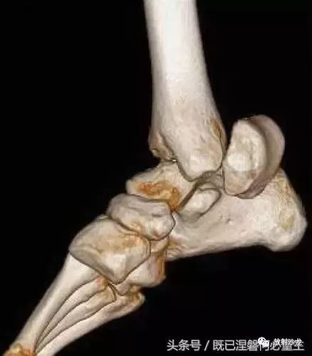
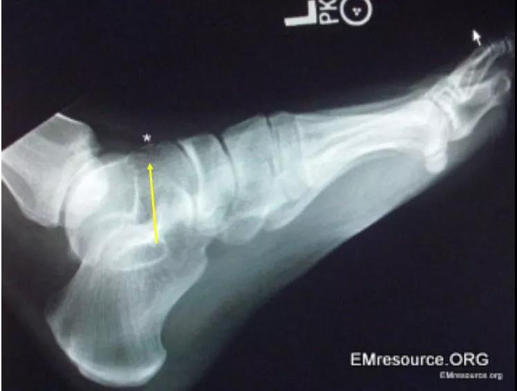
距骨侧位x线解剖图

【推荐】我自己收集总结的正常x线解剖图片学习,已更新常见x线疾病
图片尺寸450x363
膝关节解剖 下胫腓联合损伤 跟骨骨折治疗 抗生素合理应用 距骨
图片尺寸1080x810
距骨骨折和脱位:放射科医生指南
图片尺寸438x500
距骨骨折ppt
图片尺寸1080x810
踝关节正位,踝穴位,侧位及足正位,斜位↓ 关节距
图片尺寸726x628
详细的踝关节x线解剖变异解读测量
图片尺寸715x273
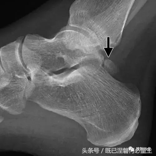
踝部的侧面x线片显示在距骨后方的椭圆形皮质骨化(箭头).
图片尺寸500x500
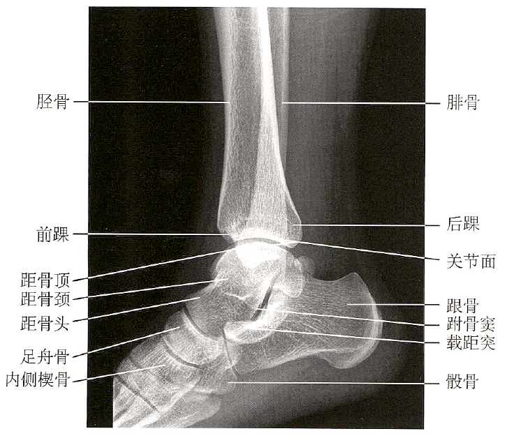
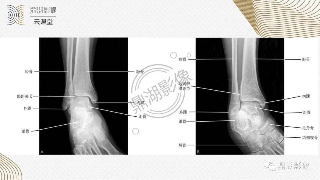
踝关节又称为距小腿关节,由腓骨远端,胫骨远端,距骨滑车组成,胫骨下端
图片尺寸524x621
在x线检查上,我们可以看到所有的骨性结构,包括胫骨,腓骨,距骨以及
图片尺寸1219x815
详细的踝关节x线解剖 变异 解读 测量
图片尺寸1898x2250
踝关节正位x线超声:可代替或结合mri检查软组织的损伤;ct:能进一步
图片尺寸632x720
跟距骨桥的微创治疗(关节镜下切除术)
图片尺寸411x322
距骨骨折和脱位放射科医生指南
图片尺寸496x500
超赞这个踝关节mri解剖课件要给满分
图片尺寸1080x608
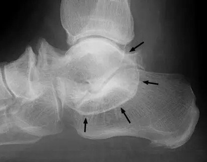
距骨骨折与脱位放射科医师及时诊断与分类指南
图片尺寸640x480
关于距骨骨折的图谱都帮你整理好了
图片尺寸748x565
日常理解的踝关节(ankle joint)是由胫骨,腓骨下端的关节面与距骨滑车
图片尺寸815x1280
距骨骨折和脱位:放射科医生指南
图片尺寸437x500
里面的主要解剖结构显示…… 第1页 你可能喜欢 正常x线解剖图谱 盆腔
图片尺寸1080x810
脱位x线片踝关节侧位片可以看到胫骨远端,腓骨远端,后踝,跟骨,距骨,足
图片尺寸601x814